The Management of Allergic Eye Diseases in Primary Eye Care
Allergy is an over-reaction of the body's immune system to innocuous foreign substances or allergens that the
body perceives as a potential threat or undesirable. It has a signi? cant impact on the quality of life of the
individual. Allergic diseases are those conditions in which an antibody- and/or T-cell mediated mechanisms are
involved. The management of allergic eye disease is aimed at preventing the release of mediators of allergy,
controlling the allergic in? ammatory cascade and preventing ocular surface damage secondary to the allergic
response. In the management of ocular allergic disease, non-pharmacologic and pharmacologic therapeutic regimens
that address the acute presentation of ocular allergy and provide prophylaxis aimed at providing long-term
maintenance therapy are advised to be recommended. This approach to the management of allergic eye diseases aims
to minimize the impact of the allergic reaction on the individual's quality of life
Allergy is an over-reaction of the body's immune system to harmless foreign substances or allergens, which
the body perceives as undesirable. It is a malady that is estimated to affect approximately 20% of the
population, of which, at least 20% suffer from ocular allergy. Allergic diseases have a significant impact on
the quality of life and wellbeing of the individual. It affects learning performance in school-aged individuals
and work productivity in adults.
Irritants or allergens such as pollen, animal dander, drugs, cosmetics, molds, and house dust mite, aromatic
hydrocarbons from diesel exhaust fumes can trigger ocular allergy. The mechanism of allergic eye disease
includes: (1) sensitization process (allergen-induced IgE production and mast cell sensitization); (2) early
phase response (allergen-induced IgE-mediated mast cell degranulation and the release of preformed mediators and
chemotactic factors); and (3) late phase response (persistent mast cell activation and TH2- mediated
DTH reaction involving the actions of newly formed mediators and chemotactic factors that incite the recruitment
of additional inflammatory cells that leads to the allergen-induced late phase allergic response).
The treatment options for management of allergy range from non- pharmacological to pharmacological management
using supportive therapy, prophylaxis and therapeutic pharmaceuticals. The adequate knowledge of the disease
process of the different types of ocular allergies along with the different classes of anti-allergic
pharmaceutical agents used in the management of these diseases will ensure success in the management of ocular
allergy and that the prescribed drug is the most appropriate for the patient's allergic eye disease.
The primary aim of a non-pharmacological approach is to prevent the onset of allergic eye reaction and/or
minimize the effect of the allergens. It is considered the first-line treatment of ocular allergy and includes
avoidance of known allergens; saline irrigation; avoid eye rubbing; and palliative therapy with cold compresses
and preservative-free ocular lubricants.
Patients should be advised to stay indoors and keep the windows closed or wear a filtermask if it is necessary
for them to stay outside on days when the pollen count is high. They should also be advised to be more aware of
the pollen count through the weather forecast; avoid cutting grass on windy days and when the pollen count is
high; and avoid going near freshly cut grass during the height of the allergy season. Furthermore, the patient
should be advised to avoid damp areas as these are potential breeding grounds for moulds; keep pets out of the
bedroom, since their hair/fur can collect allergens; and keep the kitchen and bathroom clean and dry, so as to
minimize the growth and/or breeding of mould. Also patients should be advised to keep preservative free ocular
lubricants in the refrigerator given that the cold temperature of the refrigerated preservative-free ocular
lubricants in conjunction with cool compresses induces vasoconstriction. Preservative-free ocular lubricants
dilute and flush away allergens that may come into contact with the ocular surface. They also form a barrier to
inhibit allergen contact with the ocular surface.
Pharmaceutical agents used in the management of allergic eye diseases can be classified in two main categories:
anti-inflammatory and anti-allergic therapeutic agents. The anti-allergic therapeutic agents available for the
management of acute expressions of ocular allergy and for prophylaxis are as follows:
I. Antihistamine/Vasoconstrictor Combinations
Antihistamine/vasoconstrictor combinations contain both an antihistamine (e.g. antazoline phosphate) and
vasoconstrictor (e.g. naphazoline or phenylephrine). The antihistamines in these drops help to decrease the itch
associated with allergic conjunctivitis. The vasoconstrictors in these agents are adrenergic agonists that
stimulate α-adrenoreceptor, and as such are capable of reducing the chemosis, redness and eyelid edema in
allergic eye diseases that occurs as a result of tissue congestion.
Antihistamine/vasoconstrictor combination agents are indicated for relieving the symptoms and signs of allergic
conjunctivitis. These combination agents are relatively fast acting but the duration of action last only 2-4 h,
so patients may need to dose more frequently, which may cause ocular adverse effects.
The adverse effects of these agents include burning and stinging on instillation; mydriasis, especially in
patients with lighter irides; follicular conjunctivitis; eczematoid blepharoconjunctivitis; and rebound
hyperemia or conjunctivitis medicamentosa with long-term use.
The nonspecificity of the pharmacologic action of antihistamine/ vasoconstrictors combination agents in dealing
with the ocular allergic response and its adverse ocular effects makes it inappropriate for long- term use.
II. Antihistamines
Antihistamines are very beneficial and effective pharmaceutical agents for treating individuals who present with
clinical expressions of IgE-mediated ocular allergic response. They prevent histamine released from mast cells
from interacting with histamine receptors by binding competitively to H1 receptors on the nerves and
blood vessels which results in reduced itching sensation and relief from conjunctival redness and swelling.
The first-generation and second-generation oral antihistamines, such as chlorpheniramine and cetirizine, are
H1 antagonists but they may lead to tear film insufficiency with consequential weakening of the
protection the tear film offers against environmental allergens and pollutants.
The new generation topical antihistamines such as levocabastine 0.05% and emedastine 0.05% are superior to the
first generation topical antihistamines and work by competitively blocking histamine receptors on the ocular
mucosal surface.
Emedastine difumarate 0.05% ophthalmic solution, a safe and effective selective histamine H1-receptor
antagonist, is indicated for the temporary relief of the signs and symptoms associated with allergic
conjunctivitis.
Levocabastine HCl ophthalmic suspension 0.05%, a cyclohexy-3- methylpiperidine derivative, is a potent topical
antihistamine suspension with rapid onset of action of 15 min and prolonged clinical effect lasting up to 4 h.
III. Antihistamine/Mast Cell Stabilizer
Antihistamine/mast cell stabilizer is a multimodal action antiallergic agent that acts as both a histamine
receptor antagonist and mast cell stabilizer. Olopatadine ophthalmic solution, Epinastine ophthalmic solution,
Ketotifen ophthalmic solution, and Azelastine ophthalmic solution are topical anti-allergic pharmaceutical
agents that are readily available.
Olopatadine HCl ophthalmic solution is a multimodal antiallergic agent that possesses selective H1-receptor
antagonism and mast-cell stabilizing features. It inhibits the release of allergic mediators from mast cells and
prevents histamine from providing its effects on the eye. It has a rapid onset of action and prolonged clinical
effect. The prolonged clinical effect of Olopatadine could be attributed to its ability to suppress the release
of allergic mediators and inhibit inflammatory cell recruitment. It reduces all signs and symptoms of allergic
conjunctivitis, including redness, itching, chemosis, tearing and eyelid edema. Olopatadine is approved for the
treatment of the signs and symptoms of allergic conjunctivitis.
Azelastine HCl 0.05% ophthalmic solution, a phthalazinone derivative, is a twice-a-day dosed multiple-action
anti-allergic agent. It has a rapid onset of action and is capable of inhibiting release of allergic mediators.
Azelastine ophthalmic solution is indicated for the treatment or relief of ocular itching associated with
allergic conjunctivitis. It has a noticeable bitter taste and causes ocular stinging upon instillation.
Ketotifen fumarate 0.025% ophthalmic solution, a benzocyclo- heptathiophene derivative, is a twice-a-day dosed
multi-modal anti- allergic agent. It possesses both mast-cell stabilizing and strong H1 receptor antagonism. It
is approved for the temporary prevention of ocular itching due to allergic conjunctivitis.
Epinastine 0.05% ophthalmic solution, an antihistamine with mast- cell stabilizing and anti-inflammatory
properties, is a twice daily dosed multimodal action anti-allergic agent. It is indicated for the prevention of
itching associated with allergic conjunctivitis. It has affinity for H1 and H2 receptors with prolonged
therapeutic effect lasting up to 12 h and onset of action within 3 min
Azelastine, Ketotifen and Epinastine are indicated for itching associated with allergic conjunctivitis, except
Olopatadine that is indicated for the relief of all signs and symptoms of allergic conjunctivitis.
IV. Mast Cell Stabilizers
Topical mast cell stabilizers prevent the release of allergic mediators from mast cells by suppressing mast cell
degranulation. The blockade of mast cell degranulation will inhibit the release of preformed and newly formed
mediators of allergy as well as chemokines and cytokines. They also achieve their clinical effect by preventing
calcium influx across the cell membrane. Mast cell stabilizers are safe and effective for the long term
therapeutic management of allergic eye diseases. They require a loading time of up to two weeks since it has no
effect on acute allergic reactions induced by histamine already released on the ocular surface.
Sodium cromoglycate is a mast cell stabilizer that is suitable for long- term prophylaxis and therapeutic
management of ocular allergy. It is not as effective at inhibiting the degranulation of mucosal mast cells and
this may explain the poor response of chronic ocular allergies to sodium cromoglycate. The major adverse effect
is burning and stinging.
Lodoxamide tromethamine 0.1% ophthalmic solution possesses mast-cell stabilizing properties that are considered
to be 2500 times more potent than that of sodium cromolyn. It is safe for use in children and adults on a
four-times-daily dosing for up to 3 months. The most common side effects are burning or stinging.
Pemirolast potassium ophthalmic solution, a pyridopyrimidine compound, is a mast cell stabilizer that is used for
the treatment of seasonal allergic and vernal conjunctivitis. It is dosed four times daily and it is safe for
use in children as young as 2 years of age.
Nedocromil sodium 2% ophthalmic solution, a pyranoquinoline dicarboxylic acid, is a potent stabilizer of both
connective and mucosal type mast cells. In patients with chronic ocular allergies, nedocromil sodium has been
shown to be more effective than sodium cromoglycate in preventing release of allergic mediators from the mucosal
type mast cell. It is safe and well tolerated for the long-term management of ocular allergies. It is associated
with ocular stinging or burning upon instillation and a distinctive taste.
V. Non-Steroidal Anti-Inflammatory Drugs
Non-steroidal anti-inflammatory drugs are safe for long-term use and act by blocking the cyclooxygenase pathway;
thus preventing the subsequent formation of prostaglandins, which are responsible for the itch associated with
allergic eye diseases.
Ketorolac tromethamine and diclofenac sodium are approved for use in seasonal allergic conjunctivitis for the
relief of ocular itching. Diclofenac sodium 0.1% is capable of controlling signs and symptoms of vernal
keratoconjunctivitis (VKC).
Ketorolac tromethamine 0.5% ophthalmic suspension, a pyrazolon, is a four times- a-day-dosed NSAID. It is
valuable as an adjunctive treatment in ocular allergic inflammation due to its potential to reduce the ocular
pruritus, edema and conjunctival hyperemia associated with ocular allergy. It does cause mild transient ocular
burning and stinging upon instillation, which could be alleviated by advising the patient to refrigerate the
agent prior to administration.
Topical NSAIDs blocks the production of prostaglandins but have no effect on other allergic inflammatory
mediators, such as histamine and leukotrienes that contribute to the ocular manifestations of allergic eye
disease. For this reason, it is likely to be of limited use in the treatment of ocular allergy.
VI. Corticosteroids
Corticosteroids are potent anti-allergic agents that are considered most appropriate for the treatment of chronic
ocular allergic conditions. They have the ability to suppress the recruitment and activation of pro-
inflammatory allergic mediators during the late phase of ocular allergic response. It blocks the action of mast
cell phospholipase A2, an enzyme necessary for the hydrolysis of arachidonic acid, thereby reducing the
production of late-phase allergic inflammatory mediators such as prostaglandins and leukotrienes.
Steroids are incapable of blocking histamine receptors. However, they can inhibit histamine production in mast
cells by blocking the action of histidine decarboxylase. Additionally, corticosteroids can reduce the amount of
unbound histamine on the ocular surface by increasing the availability of histaminases. Corticosteroids are not
effective in stabilizing mast cells due to their inability to prevent calcium influx into the mast cell.
However, they are capable of inhibiting T-lymphocyte response and production of cytokines such as interleukin
(IL)-4 and IL-5 production. Furthermore, corticosteroids can also inhibit neovascularisation and reduce
capillary permeability.
Loteprednol Etabonate ophthalmic suspension, a site-specific soft steroid, primarily targets the late phase
inflammatory aspect of the ocular allergic response. Loteprednol is more lipophilic than ketone corticosteroids
such as prednisolone, and it's highly lipophilic nature may contribute to its increased efficacy by
enhancing penetration into target inflammatory cells. The long-term use of loteprednol is most likely to have a
lower propensity to induce IOP elevation. Loteprednol is a suitable long-term therapeutic option for chronic
ocular allergies due to their superior safety profile relative to the ketone corticosteroids. Clinical studies
have shown that loteprednol etabonate is a safe treatment option for individuals with allergic ocular
inflammation. It has been demonstrated to be effective in treating allergic conjunctivitis and giant papillary
conjunctivitis (GPC).
Prednisolone acetate is an excellent choice for treating severe inflammatory disorders due to its good ocular
penetrability while prednisolone sodium phosphate may be considered a suitable drug for moderate inflammatory
disorders of the ocular surface due to its reduced ocular penetrability when compared to prednisolone acetate.
The fluorometholone-based steroids are used for treating mild to moderate ocular surface inflammatory conditions
and chronic inflammatory conditions requiring prolonged steroidal therapy. They have reduced propensity to
induce an IOP rise. The acetate form of fluorometholone-based steroids has greater anti-inflammatory activity
than the alcohol form (FML) due to their enhanced bioavailability. Clinical studies have shown that FML reduces
the clinical expression of VKC, including discharge, conjunctival redness, papillary hypertrophy and
tranta's dots.
VII. Immunomodulators
Immunomodulators, such as cyclosporine and tacrolimus, play vital roles in the treatment of allergic eye diseases
due to their ability to inhibit calcineurin, a phosphate that is essential to the FceRI-mediated release of
preformed mediators from mast cells. They are capable of blocking mast cell proliferation, inhibiting histamine
release, blocking release of cytokines from T-lymphocytes, and reducing eosinophil chemotaxis.
Tacrolimus is a potent macrolide immunosuppressor in ocular allergy. It exerts its action by inhibiting IgE
production by activated B-cells as well as inhibiting T-cell activation and associated proinflammatory cytokine
production. It also interferes with mast cell degranulation; inhibits the antigen-presenting role of Langerhans
cells and late phase responses in ocular allergy. It has been shown to be well tolerated with an excellent
safety profile for the long-term management of dermatitis and also it could be therapeutically beneficial for
patients with atopic keratoconjunctivitis (AKC), atopic eyelid disease and atopic dermatitis. Transient burning
sensation and recurrent herpetic lesions are reported side effects of tacrolimus.
Cyclosporine is a fungal anti-metabolite that inhibits T-lymphocyte activation via IL-2 by blocking
IL-2 production. It also reduces eosinophil chemotaxis via inhibition of IL-2 production.
Furthermore, it inhibits TH-2-lymphocyte proliferation and IFN-g with consequential modulating effect
on Th1- lymphocyte mediated DTH. Cyclosporine is prescribed, when steroids are contraindicated or unsafe, for
treating the severe form or steroid resistant cases of chronic ocular allergy.
In the management of ocular allergic diseases, non pharmacologic and/or pharmacologic therapeutic regimens that
address the acute presentation of ocular allergy and provide prophylaxis should be recommended. This approach to
the management of allergic eye diseases will minimize the adverse effect the allergic reaction may have on the
individual's quality of life.
Allergic conjunctivitis, a bilateral, self-limiting IgE-mast cell mediated conjunctival inflammatory process.
Seasonal allergic conjunctivitis (SAC) is an IgE-mast cell mediated hypersensitivity response to seasonal
allergens such as pollen whereas Perennial or persistent allergic conjunctivitis (PAC) is an IgE-mast cell
mediated hypersensitivity response to perennial allergens such as animal dander and dustmite.
The first line of therapy for patients with allergic conjunctivitis is to avoid the offending allergens. Mild to
moderate cases of allergic conjunctivitis will respond to antihistamine, topical multimodal anti-allergic
agents, mast cell stabilizers and supportive therapy. Individuals that present with hyperacute expressions of
allergic conjunctivitis, severe allergic conjunctivitis or allergic conjunctivitis that is refractory to
conventional anti-allergic therapy would benefit from short-term steroidal therapy aimed at suppressing the
allergic inflammatory cascade.
Patients who present with allergic rhinoconjunctivitis would benefit from using a nasal spray (e.g. antihistamine
nasal spray {Azelastine hydrochloride}) or oral antihistamine to provide therapeutic relief for the rhinitis and
a topical multimodal anti-allergic ophthalmic agent to treat the allergic conjunctivitis.
In the strict sense of it, giant papillary conjunctivitis is not a true allergic condition; rather it is a
chronic inflammatory condition that results from chronic irritation of the tarsal conjunctiva. The
pathophysiology of GPC is a combination of mechanical trauma to the tarsal conjunctiva and mast cell-
eosinophil-TH2 lymphocyte mediated immune hypersensitivity reaction. A poorly fitted contact lens can
potentially irritate the superior tarsal conjunctiva resulting in mechanically induced GPC. Immunologic GPC and
ocular surface inflammation in contact lens wearers may occur due to an immune response to denatured protein in
the biofilm on the contact lens surface. It has been shown that contact lens tends to attract proteinaceous
deposits on their surface. These proteinaceous deposits may become antigenic with the potential of developing a
hypersensitivity response. In order to prevent or minimize the buildup of potential antigenic biofilm on the
contact lens surface, the clinician should recommend precautionary measures, such as regular contact lens
cleaning with appropriate solutions and weekly enzymatic treatment.
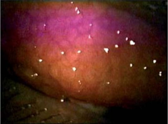
GPC is characterized by itching, tearing, burning, increased mucus production, giant papillae on the superior
tarsal conjunctiva and conjunctival injection (Fig 1). In severe cases, corneal erosion and superficial punctate
keratitis may occur. The stages include preclinical, mild, moderate and severe GPC.
The aim of management is to reduce or prevent the clinical expression of GPC by controlling, avoiding or removing
the causative agents or antigen. In case of failure of this approach, patients with GPC may benefit from either
anti-allergic therapy or pulse topical steroidal therapy depending on the degree of clinical expression of the
condition.
Patients who have GPC with significant conjunctival papillary hypertrophy or inflammatory component require both
a mast cell stabilizer and topical steroid with the intention of tapering the steroid once an adequate clinical
response has been achieved.
Contact lens wearers with allergic conjunctivitis or GPC may benefit from a topical anti-allergic agent, using
either once or twice-a-day dosing. The dose for the anti-allergic agent can be administered 5-10 min prior to
contact lens insertion and if a second dose is used, it is taken after contact lens removal at the end of the
day.
Nedocromil, a twice-a-day dosed mast cell stabilizer, may be beneficial be for long-term prophylaxis (prior to
the allergy season in seasonal allergic sufferers or all year round in perennial allergic sufferers) for contact
lens wearers who have either GPC or allergic conjunctivitis. Olopatadine 0.2% may be suitable topical multimodal
anti-allergic agent for contact lens wearers who have allergic conjunctivitis.
The most suitable course of therapy for patients with mild GPC or allergic conjunctivitis is to discontinue
contact lens wear or make several changes in their contact lens regime. The best course of management for
contact lens wearers with moderate-to-severe GPC or allergic conjunctivitis is to cease contact lens wear until
the symptoms have completely resolved.
Vernal keratoconjunctivitis is a sight-threatening, bilateral, chronic allergic inflammation of the ocular
surface that predominantly affects children and young adult males. Patients who have VKC are very symptomatic
during spring and summer and experience symptoms such as tearing, extreme itching, photophobia and mucus
discharge. In VKC, the corneal findings include superficial punctate epithelial keratitis, epithelial
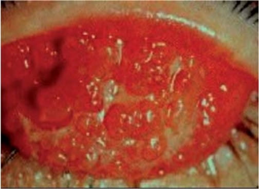
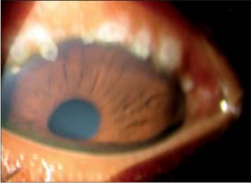
macroerosion, shield ulcer and corneal plaque. The palpebral form of VKC is characterized by giant papillae on
the upper tarsal plate while the limbal form is characterized by gelatinous limbal infiltrates (Trantas dots)
(Fig. 2 and 3).
The immunopathogenesis of VKC is a chronic IgE-mast cell and eosinophil-Th2 lymphocyte mediated allergic
inflammatory reaction with an over expression of eosinophils, Th2- lymphocytes, Th2-derived cytokines,
chemokines and other inflammatory cells or mediators. Bonini et al. proposed a clinical grading to help
physicians in the diagnosis and management of VKC as mild, moderate or severe.
The clinical manifestations of the mild form of VKC includes occasional pruritus, mild tearing, mild discomfort
including burning, stinging, foreign body sensation, mucoid discharge, mild photophobia, mild to moderate
papillary reaction, mild corneal epithelial defects with or without corneal neovascularization, subepithelial
fibrosis of the conjunctiva and mild redness and edema of the eyelid. The clinical manifestations of the
moderate form of VKC includes persistent itching, intermittent tearing, moderate discomfort, moderate mucoid
discharge with the presence of crust upon awakening, moderate photophobia, moderate tarsal papillary hypertrophy
and hyperemia with hazy view of the deep vessels, Horner-Trantas dots, moderate corneal epithelial defects with
corneal neovascularization, moderate subepithelial fibrosis and moderate eyelid inflammation with Meibomian
Gland Dysfunction. The clinical manifestations of the severe form of VKC includes constant itching and tearing,
severe discomfort, marked mucoid discharge with agglutination of the eyelids upon awakening, extreme
photophobia, severe tarsal papillary hypertrophy and hyperemia obscuring the visualization of the deep tarsal
vessels, severe corneal epithelial defects in the form of corneal erosion or ulceration with corneal
neovascularization, numerous Horner-Trantas dots, the presence of symblepharon and severe inflammation of the
eyelid.
In the management of VKC, the clinician should provide therapy that suppresses the allergic inflammatory process
before the patient becomes symptomatic. The supportive therapy of VKC includes avoiding the trigger factors such
as sun, wind, and salt water with the use of sunglasses, hat with visors and swimming goggles, avoiding eye
rubbing and applying cool compress and/or administering preservative-free ocular lubricants. Patients with a
history of VKC should be started on prophylactic therapy with mast cell stabilizers prior to the allergy season
or should remain on the therapy all year round, depending on the allergic exposure and duration of the allergic
expression.
Patients with mild VKC may benefit from supportive therapy as well as topical multimodal anti-allergic agents
and/or mast cell stabilizers to provide symptomatic relief. However, a pulse topical steroidal therapy will
become necessary to control the inflammation, if the anti-allergic therapy does not provide satisfactory
therapeutic relief.
Patients with moderate or severe VKC require concurrent use of topical steroids and mast cell stabilizers or
antihistamine/mast cell stabilizer combinations to achieve an adequate therapeutic effect. E.g.: Loteprednol
etabonate 0.5% and prednisolone acetate 0.1%. Topical cyclosporine is a suitable substitute for topical steroid
and it has been shown to be an effective long-term therapeutic agent for VKC. However, treatment with systemic
corticosteroids may become necessary when individuals present with very severe VKC that does not respond to
conventional topical therapy.
Atopic keratoconjunctivitis is a sight-threatening, bilateral, chronic allergic inflammation of the ocular
surface and the eyelids often associated with atopic dermatitis and other allergic disorders. It affects mostly
males usually in their late teens with a peak onset between the ages of 30 and 50. It is characterized by
chronic itching, burning, tearing, photophobia, papillary hypertrophy, conjunctival hyperemia, chemosis, and
thickened, erythematous, swollen eyelids. In AKC, the effect of toxic allergic mediators on the ocular surface
can cause corneal complications such as neovascularisation, pannus and corneal thinning and scarring (Fig. 4).
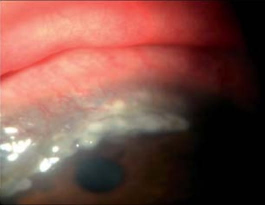
AKC is a chronic IgE-mast cell and eosinophil-T-lymphocyte mediated allergic inflammation of the ocular surface
and eyelids with an over-expression of T-lymphocytes, eosinophils, mast cells, TH1- and
Th2-derived cytokines and other inflammatory cells and mediators. Calonge et al. proposed a clinical
grading to help physicians in the diagnosis and management of AKC as Patients with mild forms of AKC (Grade I)
present with mild/occasional itching, burning and photophobia, Patients with mild-to-moderate forms of AKC
(Grade II) present with mildly persistent or moderate/occasional itching, burning and photophobia, Patients with
moderate forms of AKC (Grade III) present with moderately persistent itching, burning and photophobia and
Patients with severe forms of AKC (Grade IV) present with severe itching, burning and photophobia.
In managing individuals with AKC, focus should be on developing a management plan that will provide symptomatic
relief and prevent the development of ocular complications associated with AKC, as well as advise the patient on
the need to avoid potential trigger factors. Patient with the mild form of AKC may benefit from the use of
supportive therapy as an adjunct to topical anti-allergic pharmacologic therapy and oral antihistamine. Moderate
and severe cases of AKC associated with corneal compromise require topical steroids and prophylactic
antibiotics.
Topical immunomodulators such as cyclosporine could be used as a substitute for patients with steroid-dependent
AKC. Cool compresses and preservative- free ocular lubricants provide symptomatic relief when there is intense
ocular irritation and burning due to tear film insufficiency. The chronic blepharitis and meibomianitis
associated with AKC requires regular eyelid hygiene, warm compresses and long-term therapy with systemic
antibiotics such as doxycycline that are capable of inhibiting the action of lipase, an enzyme that converts
glandular lipids into free fatty acids. Preservative-free ocular lubricants and prophylactic antibiotics should
be considered as an essential adjunctive therapeutic strategy when therapeutic bandage contact lens are used in
managing corneal complications associated with AKC.
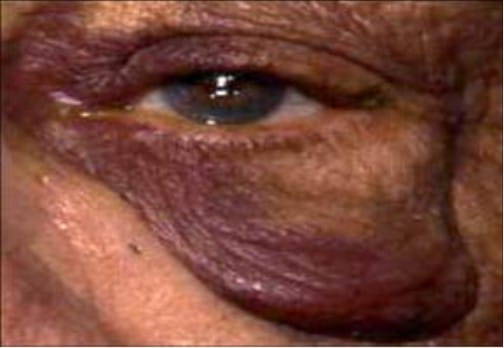
Contact Ocular Allergy is predominantly a TH1-lymphocyte mediated hypersensitivity response that
involves the ocular surface, eyelids and periocular skin. It is characterized by itching, conjunctival
hyperemia, chemosis, follicular/papillary conjunctival reaction, punctate keratitis, as well as dermatitis of
the periocular skin and eyelids (Fig. 5). Drug-induced allergic conjunctivitis (DIAC), an ocular adverse
reaction, is a hypersensitivity reaction to pharmaceutical agent applied to the eyes or periorbital region.
The management of patients with contact ocular allergy or drug-induced allergic conjunctivitis involves
identifying and removing or avoiding the offending allergen or irritant. The mild form of these conditions may
respond to palliative therapy and antihistamine, as well as, avoiding the offending allergen or irritant.
Moderate to severe cases of contact ocular allergy or DIAC require multimodal anti-allergic agents, oral
antihistamine and topical steroid to provide symptomatic relief and control of the inflammatory process.
Tacrolimus is a very effective and safe steroid sparing option for treating the periocular skin inflammation in
patients with contact ocular allergy.
It is important to understand the different types of anti-allergic and/or anti- inflammatory medications,
particularly their mode of action, the range of anti-allergic or anti-inflammatory indications, side-effect
profile, contra-indications, and relative efficacies. To ensure effective management of ocular allergies, an
inquiry about the patient's personal and family history of allergy should be done.
To promote patient compliance, anti-allergic and/or anti-inflammatory therapeutic agents that provide a rapid
clinical response and prolonged therapeutic effect should be prescribed. Also therapeutic agents should be
cost-effective, easy and convenient to use, safe for extended use, and comfortable.
The mainstay of the management of ocular allergy consists of the use of anti-allergic therapeutic agents such as
antihistamine, multiple action anti- allergic agents and mast cell stabilizers. When anti-allergic therapeutic
agents are incapable of providing adequate therapeutic relief or does not control the allergic inflammatory
process, then an anti-inflammatory agent such as topical steroid should be prescribed. Topical immunomodulators
such as tacrolimus are steroid-free alternatives that could be used when long-term use of topical steroids is
not considered a safe therapeutic option.
The management of ocular allergy depends on the severity of the allergic expression and type of ocular allergy.
To avoid ocular surface compromise and complications, accurate diagnosis and/or differential diagnosis should be
done which will guide in prescribing the most appropriate anti-allergic medication.
The management of allergic eye diseases presents a challenge to the clinician, calling for adequate knowledge of
the immunopathophysiology, clinical features and differential diagnosis of the different types of ocular
allergy. Furthermore, an adequate knowledge of anti-allergic and anti- inflammatory pharmaceutical agents used
in the management of allergic eye diseases is necessary. Knowledge of the patient's life style is helpful in
promoting compliance with recommended treatment protocols. When non-pharmacological management fails to provide
adequate relief, the next option is to prescribe anti-allergic therapeutic agents. The optimal anti- allergic
pharmaceutical agent will have a good safety profile, rapid onset of action and prolonged clinical effect.
However, pulse steroidal therapy would become necessary if the allergic expression does not respond to
anti-allergic therapy. In acute ocular allergy, the use of anti-allergic medications along with supportive
therapy is usually sufficient. In hyperacute cases and chronic ocular allergy, it is preferable to control the
inflammatory condition with an anti-inflammatory agent, such as steroids, and a prophylactic anti-allergic
agent, such as mast cell stabilizers for long-term maintenance therapy. The management of allergic eye disease
is aimed at preventing the release of mediators of allergy, controlling the allergic inflammatory cascade and
preventing ocular surface damage secondary to the allergic response. Such management can be regarded as an
essential part of primary eye care.
1. Leonardi A. Emerging drugs for ocular allergy. Expert Opin Emerg Drugs
2005;10(3):505-20.
2. Abelson MB, Leonardi A, Smith L. The mechanisms, diagnosis and treatment of allergy. Rev Ophthalmol
2002;9(4):74-84.
3. Pavesio CE, Decory HH. Treatment of ocular inflammatory conditions with loteprednol etabonate. Br J
Ophthalmol 2008;92(4):455-9.
4. Pawankar R. Inflammatory mechanisms in allergic rhinitis. Curr Opin Allergy Clin Immunol 2007; 7:1-4.
5. Reiss J, Abelson MB, George MA,Wedner HJ. Allergic conjunctivitis. In: Pepose JS, Holland GN, Wilhelmus KR,
editors. Ocular infection and immunity. St. Louis: Mosby; 1996. p. 345-58.
6. Berdy GJ. Atopic keratoconjunctivitis. Acta Ophthalmol Scand 1999;77(Suppl. 228):7-9.
7. Stahl JL, Cook EB, Barney NP, Grziano FM. Pathophysiology of ocular allergy: the roles of conjunctival mast
cells and epithelial cells. Curr Allergy Asthma Rep 2002;2(4):332-9.
8. Leonardi A. Pathophysiology of allergic conjunctivitis. Acta Ophthalmol Scand 1999;77(Suppl. 228):21-3.
9. McGill JI, Bacon A. Allergic eye disease mechanisms. Br J Ophthalmol 1998; 82:1203-14.
10. Okayama Y, Hagaman DD, Metcalfe DD. A comparison of mediators released or generated by IFN- gamma-treated
human mast cells following aggregation of Fc gamma RI or Fc epsilon RI. J Immunol 2001;166(April
(7)):4705-12.
11. Woolhiser MR, Okayama Y, Gilfillan AM, Metcalfe DD. IgG-dependent activation of human mast cells following
up-regulation of FcgammaRI by IFNgamma. Eur J Immunol 2001;31(11):3298-307.
12. Tkaczyk C, Okayama Y, Metcalfe DD, Gilfillan AM. Fcgamma receptors on mast cells: activatory and inhibitory
regulation of mediator release. Int Arch Allergy Immunol 2004;133(3):305-15.
13. Tkaczyk C, Okayama Y,Woolhiser MR, Hagaman DD, Gilfillan AM, Metcalfe DD. Activation of human mast cells
through the high affinity IgG receptor. Mol Immunol 2002;38(16-18):1289-93.
14. Chigbu DI. The pathophysiology of ocular allergy: a review. Contact Lens Anterior Eye 2009;32(1):3-15.
15. Kirkner R. Lose the CAT . . . and other pearls from allergists. Rev Optom 2001;138(3):94 8.
16. Bielory L. Ocular allergy guidelines: a practical treatment alogrithm. Drugs 2002;62(11):1611-34.
17. Abelson MB, Grant A. What to do when you see red. Rev Ophthalmol 2001;8(12):44-7.
18. Bielory L. Ocular allergy treatment. Immunol Allergy Clin North Am 2008;28(1):1-23.
19. Bielory L. Ocular allergy treatment. Immunol Allergy Clin North Am 2008;28(1):189-224.
20. Hovlin JP, DePaolis MD. Drops to prevent dropouts. Rev Optom 2000;137(Part 4):69-75.
21. Bielory L, Katelaris CH, Lightman S, Naclerio RM. Treating the ocular component of allergic
rhinoconjunctivitis and related eye disorders. MedGenMed 2007;9(3):35.
22. Butrus S, Portela R. Ocular allergy: diagnosis and treatment. Ophthalmol Clin North Am
2005;18(4):485-92.
23. Berdy GJ, Stoppel JO, Epstein AB. Comparison of the clinical efficacy and tolerability of olopatadine
hydrochloride 0.1% ophthalmic solution and loteprednol etabonate 0.2% ophthalmic suspension in the conjunctival
allergen challenge model. Clin Ther 2002;24(6):918-29.
24. Abelson MB, Gomes PJ. Olopatadine 0.2% ophthalmic solution: the first ophthalmic antiallergy agent with
once-daily dosing. Expert Opin Drug Metab Toxicol 2008;4(4):453-61.
25. D'Arienzo PA, Leonardi A, Bensch G. Randomized, double-masked, placebo controlled comparison of the
efficacy of emedastine difumarate 0.05% ophthalmic solution and ketotifen fumarate 0.025% ophthalmic solution in
the human conjunctival allergen model. Clin Ther 2002;24(3):409-16.
26. Discepola M, Deschenes J, Abelson MB. Comparison of the topical ocular antiallergic efficacy of emedastine
0.05% ophthalmic solution to ketorolac 0.5% ophthalmic solution in a clinical model of allergic conjunctivitis.
Acta Ophthalmol Scand 1999;77(Suppl. 228):43-6.
27. Noble S, McTavish D. Levocabastine an update of its pharmacology, clinical efficacy and tolerability in the
topical treatment of allergic rhinitis and conjunctivitis. Drugs 1995;50(6):1032-49.
28. Deschenes J, Discepola M, Abelson MB. Comparative evaluation of olopatadine ophthalmic solution (0.1%)
versus ketorolac ophthalmic solution (0.5%) using the provocative antigen challenge model. Acta Ophthalmol Scand
1999;77(Suppl. 228):47-52.
29. Butrus S, Greiner JV, Discepola M, Finegold I. Comparison of the clinical efficacy and comfort of
olopatadine hydrochloride 0.1% ophthalmic solution and nedocromil sodium 2% ophthalmic solution in the human
conjunctival allergen challenge model. Clin Ther 2000;22(12):1462-72.
30. Katelaris CH, Ciprandi G, Missotten L, Turner FD, Bertin D, Berdeaux G. A Comparison of the efficacy and
tolerability of olopatadine hydrochloride 0.1% ophthalmic solution and cromolyn sodium 2% ophthalmic solution in
seasonal allergic conjunctivitis. Clin Ther 2002;24(10):1561-75.
31. Abelson MB, Schwartz E. How to battle seasonal allergy. Rev Ophthalmol 1999;5(3):118-22.
32. Ciprandi G, Buscaglia S, Catrullo A, Pesce G, Fiorino N, Montagna P, et al. Azelastine eye drops reduce and
prevent allergic conjunctival reaction and exert anti-allergic activity. Clin Exp Allergy 1997;27:182-91.
33. Spangler DL, Bensch G, Berdy GJ. Evaluation of the efficacy of olopatadine hydrochloride 0.1% ophthalmic
solution and azelastine hydrochloride 0.05% ophthalmic solution in the conjunctival allergen challenge model.
Clin Ther 2002;23(8):1272-80.
34. Bielory L. Ocular allergy treatments. Clin Rev Allergy Immunol 2001;20(2): 201-13.
35. Giede C, Metzenauer P, Petzold U, Ellers-Lenz B. Comparison of azelastine eye drops with levocabastine eye
drops in the treatment of seasonal allergic conjunctivitis. Curr Med Res Opin 2000;16(3):153-63.
36. Lawrenson JG. Recent changes in the use and supply of medicines by optometrists. Optom Today
2005;45(11):28-32.
37. Greiner JV, Michaelson C, McWhirter CL, Shams NBK. Single dose of ketotifen fumarate 0.025% versus 2 weeks
of cromolyn sodium 4% for allergic conjunctivitis. Adv Ther 2002;19(4):185-93.
38. Trattler WB, Luchs J, Majmudar P. Elestat (epinastine HCl ophthalmic solution 0.05%) as a therapeutic for
allergic conjunctivitis. Int Ophthalmol Clin 2006;46(4):87-99.
39. Friedlaender MH. Epinastine in the management of ocular allergic disease. Int Ophthalmol Clin
2006;46(4):85-6.
40. Abelson MB, Smith L, Chapin M. Ocular allergic disease: mechanism, disease sub-types, treatment. Ocular Surf
2003;1(3):127-46.
41. Leonardi A. The central role of conjunctival mast cells in the pathogenesis of ocular allergy. Curr Allergy
Asthma Rep 2002;2(4):325-31.
42. Bonini S, Lambiase A, Sgrulletta R, Bonini S. Allergic chronic inflammation of the ocular surface in vernal
keratoconjunctivitis. Curr Opin Allergy Clin Immunol 2003;3(5):381-7.
43. Tauber J. Nedocromil sodium ophthalmic solution 2% twice daily in patients with allergic conjunctivitis. Adv
Ther 2002;19(2):73-84.
44. Verin PH, Dicker ID, Mortemousque B. Nedocromil sodium eye drops are more effective than sodium cromoglycate
eye drops for the long-term management of vernal keratoconjunctivitis. Clin Exp Allergy 1999;29(4):529-39.
45. Alexander M, Allegro S, Hicks A. Efficacy and acceptability of nedocromil sodium 2% and olopatadine
hydrochloride 0.1% in perennial allergic conjunctivitis. Adv Ther 2000;17(3):140-7.
46. Verin P. Treating severe eye allergy. Clin Exp Allergy 1998;28(Suppl. 6):44-8.
47. Ashrafzadeh A, Raizman MB. New modalities in the treatment of ocular allergy. Int Ophthalmol Clin J
2003;43(1):105-10.
48. Ilyas H, Slonim CB, Braswell GR, Favetta JR, Schulman M. Long-term safety of loteprednol etabonate 0.2% in
the treatment of seasonal and perennial allergic conjunctivitis. Eye Contact Lens 2004;30(1):10-3.
49. Durham SR. The inflammatory nature of allergic disease. Clin Exp Allergy 1998;28(Suppl. 6):20-4.
50. Melton R, Thomas R. Clinical guide to ophthalmic drugs. Rev Optom 2003;140(Suppl. 6):33A-9A.
51. Abelson MB, Chapin MJ. The ophthalmic drug pipeline. Rev Ophthalmol 2000;7(5):120-4.
52. Abelson MB, Hallas S, Colby K, Protopic:. A new ally against inflammation. Rev Ophthalmol
2002;9(4):104-7.
53. Nakagawa H. Comparison of the efficacy and safety of 0.1% tacrolimus ointment with topical corticosteroids
in adult patients with atopic dermatitis: review of randomised, double-blind clinical studies conducted in
Japan. Clin Drug Investig 2006;26(5):235-46.
54. Cheer SM, Plosker GL. Tacrolimus ointment. A review of its therapeutic potential as a topical therapy in
atopic dermatitis. Am J Clin Dermatol 2001;2(6):389-406.
55. Katsarou A, Armenaka M, Vosynioti V, Lagogianni E, Kalogeromitros D, Katsambas Tacrolimus ointment 0.1% in
the treatment of allergic contact eyelid dermatitis. J Eur Acad Dermatol Venereol 2009;23(4):382-7.
56. Paller AS, Eichenfield LF, Kirsner RS, Shull T, Jaracz E, Simpson EL, et al. Three times weekly tacrolimus
ointment reduces relapse in stabilized atopic dermatitis: a new paradigm for use. Pediatrics
2008;122(6):e1210-8.
57. Miyazaki D, Tominaga T, Kakimaru-Hasegawa A, Nagata Y, Hasegawa J, Inoue Y. Therapeutic effects of
tacrolimus ointment for refractory ocular surface inflammatory diseases. Ophthalmology 2008;115(6):988-92.
58. Anand MK. Immediate hypersensitivity reactions; 2004. [Retrieved on March 10, 2006 from http://www.
emedicine.com/med/topic1101.htm]
59.Brody JM, Foster CS. Vernal conjunctivitis. In: Pepose JS, Holland GN, Wilhelmus KR, editors. Ocular
infection and immunity. St. Louis: Mosby; 1996. p. 367-75.
60. Dinowitz M, Rescigno R, Bielory L. Ocular allergic disease: differential diagnosis examination techniques,
and testing. Clin Allergy Immunol 2000;15: 127-50.
61. Park AY, Scott P. IL-12: keeping cell-mediated immunity alive. Scand J Immunol 2001;53:529-32.
62. Crampton HJ. A comparison of the relative clinical efficacy of a single dose of ketotifen fumarate 0.025%
ophthalmic solution versus placebo in inhibiting the signs and symptoms of allergic rhinoconjunctivitis as
induced by conjunctival allergen challenge model. Clin Ther 2002;24(11):1800-8.
63. Lemanske Jr RF, Kakliner MA. Late phase allergic reactions. Int J Dermatol 1983;22(7):401-9.
64. Abelson MB, Rosner SA. How to handle drug-induced allergic conjunctivitis. Rev Ophthalmol
1999;6(10):146-8.
65. Donshik PC. Contact lens chemistry and giant papillary conjunctivitis. Eye Contact Lens 2003;29(Suppl.
1):S37-9 [discussion S57-9, S192-4].
66. Sindt CW, Longmuir RA. Contact lens strategies for the patient with dry eye. Ocul Surf
2007;5(4):294-307.
67. Stahl JL, Barney NP. Ocular allergic disease. Curr Opin Allergy Clin Immunol 2004; 4:455-9.
68. Ono SJ, Abelson MB. Allergic conjunctivitis: update on pathophysiology and prospects for future treatment. J
Allergy Clin Immunol 2005;115(1):118-22.
69. Siddique M, Manzouri B, Flynn TH, Ono SJ. Allergy and contact lenses. Chem Immunol Allergy
2007;92:166-75.
70. Tan ME, Demirci G, Pearce D, Jalbert I, Sankaridurg P, Willcox MD. Contact lens-induced papillary
conjunctivitis is associated with increased albumin deposits on extended wear hydrogel lenses. Adv Exp Med Biol
2002;506(Pt B): 951-5.
71. Michaud L, Giasson CJ. Overwear of contact lenses: increased severity of clinical signs as a function of
protein adsorption. Optom Vis Sci 2002;79(3):184-92.
72. Maldonado-Codina C, Efron N. Impact of manufacturing technology and material composition on the clinical
performance of hydrogel lenses. Optom Vis Sci 2004;81(6):442-54.
73. Katelaris CH. Giant papillary conjunctivitis-a review. Acta Ophthalmol Scand 1999;77(Suppl.
228):17-20.
74. Donshik PC, Ehler WH, Ballow M. Giant papillary conjunctivitis. Immunol Allergy Clin North Am
2008;28(1):83-103.
75. Abelson MB, George MA, Garofalo C. Differential diagnosis of ocular allergic disorders. Ann Allergy
1993;70(Part 2):95-109.
76. Chigbu DI. Allergic eye disease a review of the clinical features, differential diagnosis and diagnoses.
Optom Today 2006;46(15):35-8.
77. Bielory L. Differential diagnosis of conjunctivitis for clinical allergist-immunologists. Ann Allergy Asthma
Immunol 2007;98:105-15.
78. Leonardi A, Secchi AG. Vernal keratoconjunctivitis. Int Ophthalmol Clin J 2003;43(1):41-58.
79. Lemp MA. Contact lenses and allergy. Curr Opin Allergy Clin Immunol 2008;8(5):457-60.
80. Cameron JA. Shield ulcers and plaques of the cornea in vernal keratoconjunctivitis. Ophthalmology
1995;102(6):985-93.
81. Buckley RJ, Bacon AS. Giant papillary conjunctivitis. In: Pepose JS, Holland GN, Wilhelmus KR, editors.
Ocular infection and immunity. St. Louis: Mosby; 1996. p. 359-66.
82. Zhan H, Smith L, Calder V, Buckley R, Lightman S. Clinical and immunological features of atopic
keratoconjunctivitis. Int Ophthalmol Clin J 2003;43(1):59-72.
83. Nishida T, Taylor AW. Specific aqueous humour factors induce activation of regulatory T cells. Invest
Ophthalmol Visual Sci 1999;40(10):2268-74.
84. Berdy GJ. Atopic Keratoconjunctivitis. Acta Ophthalmol Scand 1999;77(Suppl. 228):7-9.
85. Buckley RJ. Allergic eye disease-a clinical challenge. Clin Exp Allergy 1998; 28:39-43.
86. Casey R, Abelson MB. Atopic keratoconjunctivitis. Int Ophthalmol Clin 1997;37(2):111-7.
87. Schmid KL, Schmid LM. Ocular allergy: causes and therapeutic options. Clin Exp Optom
2000;83(5):257-70.
88. Kapp A, Allen BR, Reitamo S. Atopic dermatitis management with tacrolimus ointment (Protopic). J Dermatolog
Treat 2003;14(Suppl. 1):5-16.
89. Lemp MA, Bielory L. Contact lenses and associated anterior segment disorders: dry eye disease, blepharitis,
and allergy. Immunol Allergy Clin North Am 2008;28(1):105-17.
90. Elher WH, Donshik PC. Giant papillary conjunctivitis. Curr Opin Allergy Clin Immunol 2008;8:445-9.
91. Donshik PC. Giant papillary conjunctivitis. Trans Am Ophthalmol Soc 1994; 92:687-744.
92. Porazinski AD, Donshik PC. Giant papillary conjunctivitis in frequent replacement contact lens wearers: a
retrospective study. CLAO J 1999; 25(3):142-7.
93. Kymionis GD, Goldman D, Ide T, Yoo SH. Tacrolimus ointment 0.03% in the eye for treatment of giant papillary
conjunctivitis. Cornea 2008;27(2):228-9.
94. Foulks GN, Harvey T, Raj CV. Therapeutic contact lenses: the role of high-Dk lenses Ophthalmol Clin North Am
2003;16(3):455-61.
95. Chigbu DI. Allergic eye disease a review of their management. Optom Today 2006; 46(16):35-8.
96. Bonini S, Sacchetti M, Mantelli F, Lambiase A. Clinical grading of vernal keratoconjunctivitis. Curr Opin
Allergy Clin Immunol 2007; 7(5):436-41.
97. Calonge M, Herreras JM. Clinical grading of atopic keratoconjunctivitis. Curr Opin Allergy Clin Immunol
2007;7(5):442-5.
98. Keklikci U, Soker SI, Sakalar YB, Unlu K, Ozekinci S, Tunik S. Efficacy of topical cyclosporin A 0.05% in
conjunctival impression cytology specimens and clinical findings of severe vernal keratoconjunctivitis in
children. Jpn J Ophthalmol 2008; 52(5):357-62
99. Adapted from: Cont Lens Anterior Eye. 2009 Dec; 32(6): 260-72