

Surgical Insights- Complications of Phacoemulsification and Manual SICS

Introduction
For a number of years, phacoemulsification has been the method of choice for cataract extraction in developed countries. Phacoemulsification can offer a safe and elegant disassembly and aspiration of the lens and a rapid recovery for patients.1
Phacoemulsification, however, is difficult to employ in high volume in developing countries as the technology requires costly machinery and consumables, a permanent and reliable source of electricity, regular maintenance, and specially trained surgeons and support staff. Phacoemulsification can also potentially lead to more serious complications when used to remove extremely dense cataracts commonly encountered in developing countries.2
Given these challenges, manual small-incision cataract surgery (SICS) has emerged as a cost-effective alternative to phacoemulsification in the developing world.3 With suture-less manual SICS, the undivided nucleus is extracted and a poly (methyl methacrylate) (PMMA) intraocular lens (IOL) is implanted through a self-sealing scleral pocket incision. This technique is less expensive in terms of capital equipment investment, equipment maintenance, and disposable costs per case. In addition, it may be faster and better suited for the advanced and mature cataracts.4
Phacoemulsification vs. Manual SICS
Gogate et al. compared phacoemulsification with manual SICS in a randomized controlled trial of 400 eyes in India and concluded that the techniques were comparable in efficacy and safety.
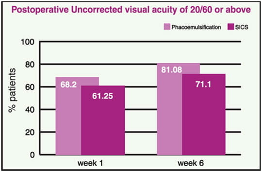
- Best corrected visual acuity of 20/60 or above at postoperative week 6 was achieved by 98.4% phacoemulsification vs. 98.4% SICS.
- Complication rates were rare and similar between both the groups & included posterior capsular opacity, iridodialysis and iritis.2
- Manual SICS was shown to be significantly faster than phacoemulsification. The average operative times plus turnover reported were 15 min 30s for phacoemulsification, and 8-9 minutes for manual SICS.2
A recent retrospective cohort series, carried out in Arvind Eye Hospital India, analysed the rate of intraoperative complications with phacoemulsification and manual SICS during a 12 month study period4. Results were as follows:
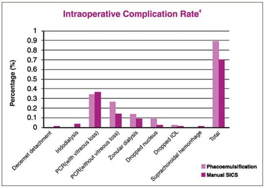
- Overall intraoperative complication rate: Phacoemulsification (1.11%), manual SICS (1.01%), ECCE (2.60%).4 Posterior capsule rupture (PCR) was the most frequent complication accounting for two thirds (67%) of all complications where vitreous loss occurred in 72% of these eyes.
- Manual SICS had a lower risk for complications than phacoemulsification among staff surgeons as well as trainees. However, overall outcomes were statistically better with phacoemulsification (96%) than with manual SICS (89%).4 The findings suggested that compared with phacoemulsification, manual SICS is comparably safe for experienced surgeons as well as less experienced cataract surgeons.4
However another retrospective cohort series carried out in L. V. Prasad eye institute evaluated outcomes of SICS and phacoemulsification when performed by trainees.5 The overall complications were seen more in the SICS group as compared with the phacoemulsification group (15.1% vs. 7.1%). Although the complication rate was higher in the MSICS group, there was no difference in BCVA in both the groups.5
In a study of more than 42,000 consecutive cataract surgeries, the rate of infectious endophthalmitis was lower after phacoemulsification than after manual SICS.3
However, with SICS, there is more risk of astigmatism if the wound construction isn’t properly done. There is also more risk of corneal oedema on the first postoperative day than with phacoemulsification. Posterior capsular opacification could be a greater risk in SICS than in the phacoemulsification. Iris injury is also slightly more common with SICS.1
Postoperative Complications of Manual SICS
Most of the postoperative complications with manual SICS are similar to those encountered with phacoemulsification.
- Immediate Complications
Wound leak, corneal complications (DM stripping), corneal endothelial damage), postoperative iritis, postoperative IOP rise.
- Late Complications
Corneal decompensation, uveitis, capsular bag complication, IOL malposition, PCO, CME, endophthalmitis.6
Management of Complications
Intraoperative Complications
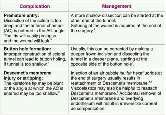
During Construction of a Self-Sealing Wound
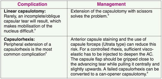
During Hydrodissection

During Nucleus Delivery
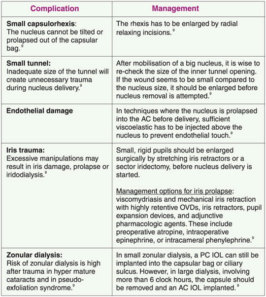
PCR may occur during hydrodissection, nucleus delivery or cleaning of cortex.9
Management: Once a PCR is noticed, irrigation should be stopped and vitreous integrity should be checked. If the anterior vitreous face is not disturbed, remaining lens cortex can be aspirated, using as little irrigation as possible. In case of any vitreous disturbance, anterior vitrectomy has to be done.9
Common Postoperative Complications
Post-Operative Corneal Oedema
Postoperative corneal oedema may occur for a variety of reasons. Toxic injury may occur to the endothelium from intraoperative medications, surgical trauma, detachment of Descemet’s membrane, inflammation, low preoperative cell count, or the Brown-McLean syndrome. Treatment of postoperative corneal oedema can include hypertonic saline and topical anti-inflammatory therapy. If endothelial cell damage is so severe that corneal edema persists, then penetrating keratoplasty may be necessary.9
Standard treatment of PCO consists of opening capsule with Nd:YAG laser. Changes in surgical techniques, as well as adjunctive pharmacologic agents, may reduce incidence of posterior capsular opacification. In opacification for congenital cataracts, primary posterior capsulectomy & anterior vitrectomy reduces its incidence9.
Incision location, size & configuration all significantly affect surgical astigmatism. Astigmatism could be decreased by: decreasing incision size & adjusting suture depth, suture material & suture size. An incision that is posteriorly located is associated with less astigmatism than an anterior wound. A straight or frown-shaped incision has less astigmatism than a curved incision parallel to limbus. If intraoperative techniques are not successful, postoperative management can include the following8
- Clinical management: Spectacles, contact lenses
- Surgical management: Astigmatic keratotomy/Corneal relaxing incisions, Limbal relaxing incisions
Endophthalmitis
The management strategy will then be directed according to the presenting visual acuity in the affected eye.
- When Visual Acuity is Better Than Light Perception
Aqueous and vitreous samples should be obtained with both an aqueous tap and either a vitreous tap or a vitreous biopsy (with a cutting and aspiration probe).10 Samples are sent for Gram stain, culture, and sensitivity. Empiric antibiotics are then administered, usually in the form of a single intravitreal injection of vancomycin and ceftazidime.11 Patients should be closely followed up and if no clinical improvement is seen within 48 hours, a repeat injection might be warranted (although the risk of retinal toxicity should be considered). 11,11
- In patients with Light Perception Vision
3-port pars plana vitrectomy should be done immediately because this regimen has demonstrated a 3-fold increase in the likelihood of 20/40 final acuity. Vitrectomy not only allows for collection of samples, but also has other benefits, including improvement of antibiotic distribution and reduction of infecting organisms, toxins, and opacities. 12,11
- In diabetic patients
Greater likelihood of achieving a 20/40 acuity with vitrectomy (57%) than with a simple tap or biopsy (40%)11
- Antibiotics are administered at the time of surgery. The prognosis of PE is variable and depends on the type of organism isolated as well as the degree of inflammatory damage to the eye. Outcomes range from excellent visual acuity to blindness and even loss of the eye altogether.
Conclusion
Treating cataract blindness worldwide continues to be a formidable challenge. Significant barriers include cost, lack of population awareness, shortage of trained personnel and poor surgical outcomes. Both phacoemulsification and manual SICS achieves excellent visual outcomes with low complication rates, but manual SICS is significantly faster, less expensive and requires less technology. Therefore, manual SICS may be the preferred technique for cataract surgery in the developing world.1
References
- Eyenet April 2009:43-47
- Curr Opin Ophthalmol 2008;19:55–59
- J Cataract Refract Surg 2010; 36:1849–1854
- J Cataract Refract Surg 2012; 38:1360–1369
- BMJ Open 2012;2:e001035.doi:10.1136/bmjopen-2012-001035 Last accessed on 9th July 2013.
- http: // www .aravind.org/sics/chapter5_7.htm Last accessed on 9th July 2013.
- Community Eye Health 2003; 16 (48):58-60.
- Indian Journal of Ophthalmol 2009;57: 45-9.
- Surv Ophthalmol 1999; 44: 123–147.
- Arch Ophthalmol 1995;113(12):1479-96
- Retina 2007;27(6):662-80
- J Cataract Refract Surg 2011; 37:1699–1714