

Hypertension and Eye-Issue 2

Introduction
Systemic hypertension is a common condition associated with significant morbidity and mortality.It confers cardiovascular risks by causing target – organ damage that includes retinopathy in addition to heart disease, stroke, renal insufficiency and peripheral vascular disease. It also causes worsening of microvascular eye disease like diabetic retinopathy if poorly controlled.1Thisissue of hypertension and eye booklet aims to provide information on the profound effects of hypertension on eye.
Impact of Hypertension on Eye
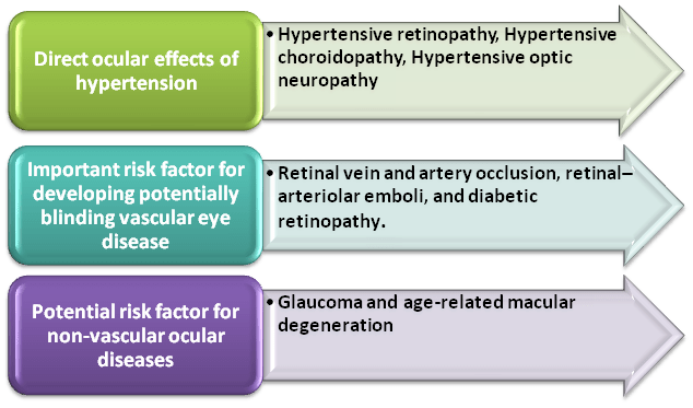
The profound effects of hypertension on eye have been classified as shown in figure 12:
Figure 1. Classification of the profound effects of hypertension on eye
Direct Ocular Effects of Hypertension2
Hypertensive Retinopathy
- It refers to retinal microvascular signs that are related to raised bloodpressure.
Prevalence
- Population-based studies detected signs of hypertensive retinopathy in 2–14% of the non-diabetic population aged 40 years and older.
Signs & Symptoms
- A 3 grade classification system has been proposed for signs of hypertensive retinopathy:
|
Mild retinopathy |
Would be identified by retinal–arteriolar signs, such as generalised and focal arteriolar narrowing, arteriolar wall opacification, and arteriovenous nipping. |
|
Moderate retinopathy |
Would be recognised by flame-shaped or blot-shaped haemorrhages, cotton-wool spots, hard exudates, microaneurysms, or a combination of all of these factors. |
|
Severe retinopathy |
Would display some or all of these retinopathy signs, as well as swelling of the optic disc |
Figure 2: (A) Signs of mild hypertensive retinopathy in an eye with ischaemic optic neuropathy. (B) Signs of severe hypertensive retinopathy
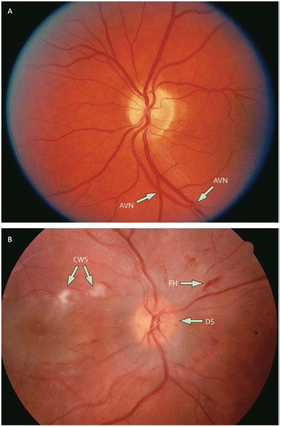
CWS=cotton-wool spots. FH=flame-shaped retina haemorrhage. DS=swelling of the optic disc. AVN=arteriovenous nipping.
- These signs were strongly associated with high blood pressure.
- Hypertensive retinopathy has long been regarded as a marker of systemic vascular disease elsewhere in the body.
- In a 3-year population-based cohort study of atherosclerosis risk, incident stroke events were more common in participants with signs of hypertensive retinopathy than in participants without retinopathy.
- Studies have now linked signs of hypertensive retinopathy with cognitive decline, cerebral white-matter lesions identified by cerebral MRI, lacunar infarctions, cerebral atrophy and stroke mortality.
- Some investigators suggest that moderate hypertensive retinopathy could be used to predict incident congestive heart failure, even in individuals without a previous history of myocardial infarction
Management
- Various national guidelines for management of hypertension recommend assessment of retinopathy to enable risk stratification.
Table 1: Retinopathy treatment based on severity
|
Severity |
Treatment |
|
Mild retinopathy |
Will probably only need routine care. |
|
Moderate signs |
Might benefit from further assessment of blood-pressure control (eg, home or 24-hour blood-pressure monitoring), assessment of other vascular risk (eg, cholesterol levels) and, if clinically indicated, appropriate risk-reduction therapy (eg, cholesterol-lowering agents). |
|
Severe retinopathy |
Need urgent antihypertensive management. |
|
Patients with borderline or so-called white coat hypertension |
Physicians could interpret mild or moderate signs of retinopathy as evidence for end-organ damage, and as an indication that antihypertensive therapy could aid in treatment. In patients with established hypertension, signs of retinopathy could suggest a need for close observation of blood pressure, supplementary antihypertensive therapy, or both. |
- Evidence suggests treatment of hypertension could reverse the changes seen with retinopathy.
Hypertension as a Risk Factor in Ocular Disease2
Retinal Vein Occlusion (RVO)
- It ischaracterised clinically by dilated and tortuous retinalveins and the presence of retinal haemorrhages,cotton-wool spots, and oedema of the macula and optic disc.
- Central retinal vein occlusion (CRVO) occurs in both ischaemic and non-ischaemic forms. Patients with an ischaemic CRVO typically present with poor visual acuity and a relative afferent papillary defect.
- Fluoroscein angiography of the fundus can show capillary non-perfusion. These patients have a poorer visual prognosis and are at risk of secondary neovascular glaucoma.
Prevalence
- Population-based surveys generally indicate that CRVOs arise in 0·1–0·4% and branch retinal vein occlusions(BRVOs) in 0·6–1·1% of adults aged 40 years and older.Participants withhypertension were 5 times more likely to have a BRVO than those without hypertension.
Risk factors
- RVO is also associated with other cardiovascular risk factors, including diabetes, cigarette smoking, carotid artery disease, and various haematological abnormalities (eg, hyperhomocysteinaemia, anticardiolipin antibodies, protein S and C deficiencies, activated protein C resistance, and factor V Leiden mutation), coronary heart disease and cardiovascular mortality.
Management
- Management include assessment of blood pressure control, standard cardiovascular risk factors, and haematological function.
- Ophthalmic follow-up is needed to diagnose and prevent the two main complications of retinal veinocclusion: neovascularisation and macular oedema.
- Randomised clinical trials have shown that prophylacticpanretinal laser treatment does not necessarily preventneovascularisation in ischaemic vein occlusions, and thatlaser treatment can be withheld unless the patient developsrank ocular neovascularisation.
- Focal laser treatmentcan assist, however, in prevention of visual loss in somepatients with macular oedema from BRVO,but does not seem to benefit macular oedemaassociated with CRVO. Severaltreatment strategies for macular oedema (eg, steroid injections or antivascular endothelial growth factor agentsinto the vitreous) have been proposed, but their effectiveness and safety will need to be confirmed byrandomised clinical trials.
- Hypertension treatment has not been proven to reduce the risk ofcomplications associated with retinal vein occlusion, orprevent the development of this disorder in the unaffected eye, physicians should more closely monitor blood pressure and consider initiation or modification of therapy in patients with this eye disorder.
Figure 3: (A) Central retinal vein occlusion. (B) Branch retinal vein occlusion
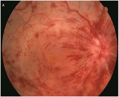
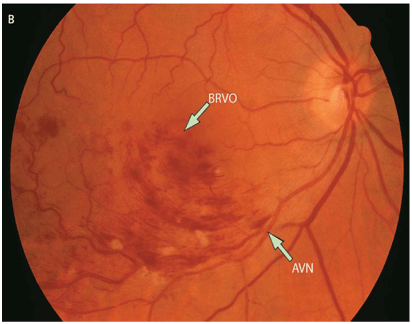
AVN=arteriovenous nipping. BRVO=branch retinal vein occlusion.
Retinal Emboli
- Retinal–arteriolar emboli are discrete plaque-like lesions, lodged in the lumen of retinal arterioles. These are heterogeneous, and can be composed of cholesterol crystals (reflective emboli) or fibrin, platelets, calcium, or other materials (non-reflective emboli).
- It can be single or multiple, and can be seen in one or both eyes.
Figure 4: Retinal–arteriolar emboli and retina branch artery occlusion
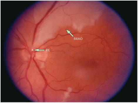
RE=retinal emboli. BRAO=branch retinal artery occlusion.
Prevalence
- Epidemiological studies report that asymptomatic retinal emboli are fairly common in adults aged 40 years and older. Two large population-based studies have reported prevalence rates of 1·3% and 1·4%.
Risk factors
- The main risk factors are hypertension, diabetes and cigarette smoking.
- Retinal emboli have two important clinical implications-
- First, the distal portions of occluded arterioles could be ischaemic, and thus, could result in frank retinal artery occlusion.
- Second, people with retinal emboli have a higher risk of thromboembolic stroke and cardiovascular disease.
Management
- Because of their increased risk of cardiovascular disease, patients with retinal emboli will need thorough systemic assessment, concentrating on hypertension control and other modifiable vascular risk factors.
- Patients with retinal emboli and atrial fibrillation will need systemic anticoagulation treatment.
Retinal Artery Occlusion
- Retinal artery occlusion occurs commonly in patients with hypertension.
- Central retinal artery occlusion presents with a sudden, painless, unilateral vision loss and typically appears as a cherry red spot.
- Occlusion of a branch retinal artery could present with a visual-field defect, and loss of central vision can be slight.
- In up to 70% of cases of branch retinal artery occlusion retinal emboli is visiblein the vessels at the optic disc, or downstream in branch retinal arterioles;these signs are present in about 20% of cases when the occlusion arises centrally.
Figure 5: Central retinal artery occlusion
Prevalence
- Clinic outpatient data estimated, the yearly incidence of central retinal artery occlusion to about one in 10000, occurring typically in people aged 60–65 years. However, a population-based study showed a significantly lower incidence of only 0·07 per 10000 people per year.
Risk Factors
- Retinal artery occlusion is associated with hypertension and other cardiovascular risk factors, with haematological abnormalities, and with both subclinical and clinical stroke.
Management
- Thorough cardiovascular and cerebrovascular assessments, including analysis of carotid and cardiac images,are necessary for patients who present with retinal artery occlusions.
- Central retinal artery occlusion is usually regarded as an ocular emergency.
- Attempts to restore ocular circulation and preserve vision include-
- Rapid dislodgement of the embolus by digital massage of the eyeball.
- Paracentesis to remove anterior chamber fluid and lower intraocular pressure
- Breathing into a paper bag to induce carbon-dioxide-related vasodilation.
- More aggressive treatment strategies such as selective ophthalmic artery fibrinolysis via the femoral artery have been suggested, but their effectiveness has yet to be proved.
Retinal Macroaneurysm
- Retinal arterial macroaneurysm, a fusiform or secular dilation of the retinal arterioles, is an uncommon disorder almost always seen in patients with hypertension.
Prevalence
- Data from large case series suggest that about a fifth of macroaneurysms are bilateral, and one in ten are multiple. It is usually an incidental finding in asymptomatic patients, but can also present acutely, with visual loss secondary to haemorrhage or exudation.
Risk Factors
- Hypertensive patients, with impaired auto regulation, are at particular risk.
- Hypertension has been reported in up to 75% of patients with macroaneurysms.Patients with uncontrolled hypertension might initially present with visual loss caused by macroaneurysm.
Management-
- Visual recovery typically occurs spontaneously with thrombosis of the macroaneurysm and resolution of the haemorrhage and exudate.
- However, residual retinal damage from chronic macular oedema and hard exudate deposition might lead to persistent poor vision.
- Anecdotal data suggest thatlaser treatment could be useful in some cases, especially when exudation affects the macula.
Ischaemic Optic Neuropathy
- Ischaemic optic neuropathy is the mostfrequent acute optic neuropathy in patients aged over50 years.
Types of Optic Neuropathy
- Either the anterior or the posterior segment of optic nerve can be affected.
- Anterior ischaemic opticneuropathy accounts for 90% of cases, and typically presents with sudden visual loss and optic-disc oedema, which is typically absent in posterior ischaemic optic neuropathy.
- Anterior ischaemic optic neuropathy can be further subdivided into 2 types-
- Arteritic- This form is typically due to giant-cell temporal arteritis, which is not associated with hypertension.
- Non-arteritic - Has been strongly linked with hypertension and other cardiovascular risk factors. Clinical series show that up to 50% of patients with non-arteritic anterior ischaemic optic neuropathy might have hypertension and 25% might have diabetes. Apart from hypertension and diabetes, hypercholesterolaemia seem to increase the risk of anterior ischaemic optic neuropathy in younger patients more than they do in older patients.
- Non-arteritic anterior ischaemic optic neuropathy has no known effective treatment.
- Visual recovery after non-arteritic anterior ischaemic optic neuropathy is often limited, but spontaneous improvement of vision did occur in patients during the first year of this trial.
Diabetic Retinopathy
- Diabetic retinopathy is the most specific microvascularcomplication of diabetes and one of the main causes ofvisual impairment, especially in people of working-age.
Prevalence
- A population-based study in the USA suggested that 33% ofdiabetic people aged 40 years and older have retinopathy,and 8% have vision-threatening retinopathy.
Risk Factors
- Raised blood pressure is anindependent risk factor for both the initial development ofretinopathy and its subsequent progression.
- Impairedretinal–vascular autoregulation in response to high bloodpressure plays a part in this association, since diabeticpatients with hypertension seem to be less able to regulate retinal blood flow than non-diabetic patients.
- In diabetes,hypertension can also result in endothelial damage in theretinal vasculature and increased expression ofvascular–endothelial growth factors.
Management
- In general, data from epidemiological studies andclinical trials lend support to clinical recommendationsthat control of hypertension and blood pressure inpatients with type 2 diabetes should help to preventretinopathy and other microvascular complications.
- Arandomised controlled clinical trial showed that intensiveblood pressure control was more beneficial than conventionalcontrol for normotensive patients with type 2diabetes but not for hypertensive patients.
- Study suggested that ACE (Angiotensin converting enzyme)inhibitors might have an additional beneficial effect inprevention of retinopathy—independent of reduction ofblood pressure.
Ocular Diseases where Hypertension is a Potential Risk Factor2,3
Age-related Macular Degeneration
- Age-related macular degeneration is the most common cause of visual impairment in patients aged 65 years and older in developed countries.
- Visual loss from age-related macular degeneration typically results from either neovascularisation associated with choroidal vessels (commonly termed wet or exudative age-related macular degeneration) or geographic atrophy of the retina.
Risk Factors
- Some suggest hypertension could increase the potential risk factor for age-related macular degeneration, on the basis of its purported effects on the choroidal circulation.
- The Beaver Dam Eye Study, reported that raised SBP at baseline was not related to prevalent age-related macular degeneration, but did increase the 10-year risk of the disorder.
- Many of the risk factors for cardiovascular disease (such as cigarette smoking, carotid artery disease, and systemic markers of inflammation) also predispose patients to this disorder.
- Furthermore, the disorder has been linked with a high risk of stroke and cardiovascular mortality.
Management
- A wide range of treatment options for age-relatedmacular degeneration, including vascular endothelialgrowth-factor inhibitors, have been developed in the pastdecade. However, specific antihypertensive medicationor treatments to lower blood pressure have not proven beneficial for prevention of the development or progressionof disorder. Observational studies suggest thatantihypertensive medications do not affect the risk of thisdisorder.
Glaucoma
- Glaucoma is a group of disorders characterised by progressive damage to the optic nerve and visual field loss.
Prevalence4
- A recent study on Indian glaucoma patients has shown that 47.5% of glaucoma patients suffered from hypertension.
Risk Factors
- Although certainly not the only risk factor, there is adequate evidence to show that abnormalities in blood pressure and blood flow play a central role in glaucoma pathogenesis.
- In particular, low blood pressure predisposes to low ocular perfusion pressure (OPP) increasing the likelihood of hypoxic or ischaemic stress.This is pertinent in that nocturnal IOP elevations and blood pressure dips can act synergistically to produce substantial OPP troughs over a diurnal cycle, which have been implicated in the development of normal tension glaucoma.
Management
- An epidemiological study has shown that overtreatment of hypertension increases glaucoma risk.The effect of high blood pressure is more complex.
- In the short term,high blood pressure can improve the OPP and provide some protection against IOP induced ischaemia whilethe influence of chronic presentations on glaucoma remains controversial among epidemiological studies and is not well established in animal experiments.
- The most recent evidence from an epidemiological trial showed that hypertension predisposes to the development of glaucoma. This is in line with the widespread vascular damage frequently associated with chronic hypertension, which would act to impair ocular blood flow and autoregulation (secondary vascular dysregulation).
- The presence of impaired autoregulation means that the eye is less able to cope with episodes of low OPP and over time a cumulative effect could produce ganglion cell loss. In an attempt to adapt to chronic high blood pressure, a rightward shift of the cerebral blood flow autoregulatory curve has been found in patients with hypertension and animal models.
- If this can be extrapolated to the eye, it provides an explanation of why patients with hypertension are more vulnerable to low OPP.
- From a clinical point of view, it is important to consider not only the IOP but also the blood pressure status in glaucoma patients. Specifically, it is important to avoid under- or overtreatment of chronic hypertension to achieve an optimal OPP range.
- A study in Indian glaucoma patients reported that4-
- The most common systemic medication for hypertension was amlodipine, a calcium channel blocker (CCB), atenolol, a systemic ?-blocker.
- Almost 10%of subjects were on combined systemic and topical ?-blocker therapy.
References
- Journal of human hypertension.2002;16:6667-675
- Lancet. 2007; 369: 425–35
- ClinExpOptom. 2011; 94(2): 133–149
- Int Ophthalmol. 2013; 33:527–532