Eosinophilic Esophagitis

calendar
3 Oct, 12

Eosinophillic Esophagitis
 

Introduction

Eosinophilic esophagitis (EoE) is a condition that is recognized by its symptoms, histological and endoscopic features. Recent interest in this disease as a separate clinical entity is perhaps the reason for increased recognition of it in adults.

Previous guidelines for the diagnosis and treatment of this condition mandated fulfilling histologic and clinical criteria; however, they did not have proven biological significance or the power to discriminate amongst other esophageal diseases, leading to few physicians adopting them. The amount of research has doubled since the last guidelines on this condition were published, providing new insights into this clinical entity.

The clinical demand for a more relevant diagnostic guideline lead to the development of these recommendations. The recommendations and information in this guideline were based on the literature review and the opinions of a panel of experts. Areas of EoE such as diagnostics, genetics, allergy testing, therapeutics and disease complications have been covered.

This article is a concise compilation of these guidelines, created to act as a quick guide.
Diseases associated with esophageal eosinophilia

  • Gastroesophageal reflux diseade (GRED)
  • EoE
  • Eosinophilic gastrointestinal diseases
  • Celiac disease
  • Crohn's disease
  • Infection
  • Hyper-eosinophilic syndrome
  • Achalasia
  • Drug Hypesensitivity
  • Vasculitis
  • Pemphigoid vegetans
  • Connective tissue disease
  • Graft-versus-host disease

Definition

EoE represents a chronic, immune/antigen-mediated esophageal disease characterized clinically by symptoms related to esophageal dysfunction and histologically by eosinophil-predominant inflammation.

History and Physical Examination

  • Patients should have their medical history carefully checked, and undergo a careful examination, focusing on eating and swallowing habits if they have symptoms suggestive of EoE.
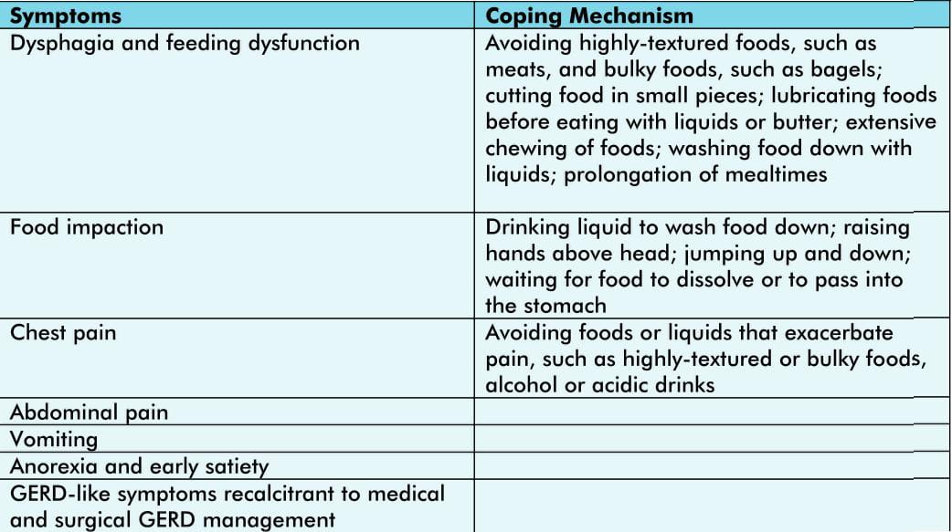
  • A high index of suspicion is necessary to detect impaired esophageal function, often camouflaged by coping behaviors (Table 1).

Table 1: Symptoms associated with EoE

CPM0GHOP03882

  • A physical examination is necessary in children to assess if the disease is affecting growth and nutrition, e.g., feeding difficulties or severe dietary restriction.
  • Appropriate evaluations should be undertaken when signs indicative of other conditions that might involve the esophagus (e.g., Crohn's disease and eosinophilic gastroenteritis) or that might mimic the condition (e.g., GERD and achalasia) are present.
  • Proton-pump inhibitor (PPI) responsiveness or diagnostic testing might not adequately distinguish GERD and EoE.

Endoscopic and Radiologic Features

In children, diagnosis of EoE only through symptoms is not feasible as the manifestationsare non-specific and vary with age.

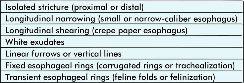
  • Endoscopy with esophageal biopsy remains the only reliable diagnostic test for EoE.
  • Isolated esophageal eosinophilia without associated symptoms and without ruling out other causes of esophageal eosinophilia is inadequate to make the diagnosis of EoE.
  • In patients with unexplained dysphagia, an esophageal biopsy specimen should be taken to seek histologic evidence of EoE even if the endoscopy results appear normal. This should be done to identify a potential cause of dysphagia other than EoE.
  • Two to four mucosal biopsy specimens of the proximal and distal esophagus should be obtained.
  • In children and adults (when indicated), biopsy specimens of the gastric antrum and duodenum should be obtained once to exclude other causes of esophageal eosinophilia.
  • Although it is reasonable to perform gastric or duodenal biopsies, there is limited evidence to recommend routine biopsies in the absence of symptoms or endosopic abnormalities suggesting other gastrointestinal disorders.
  • Radiography is useful only to characterize anatomic abnormalities and is not recommended as a routine diagnostic test for EoE.
  • Food impaction necessitating endoscopic bolus removal occurs in 33-54% of adult with EoE.

Table 2: Endoscopic and radiologic features of EoE

CPM0GHOP03885

Histologic Findings

  • Histopathologic features of esophageal mucosal biopsy specimens must be interpreted along with clinical information.
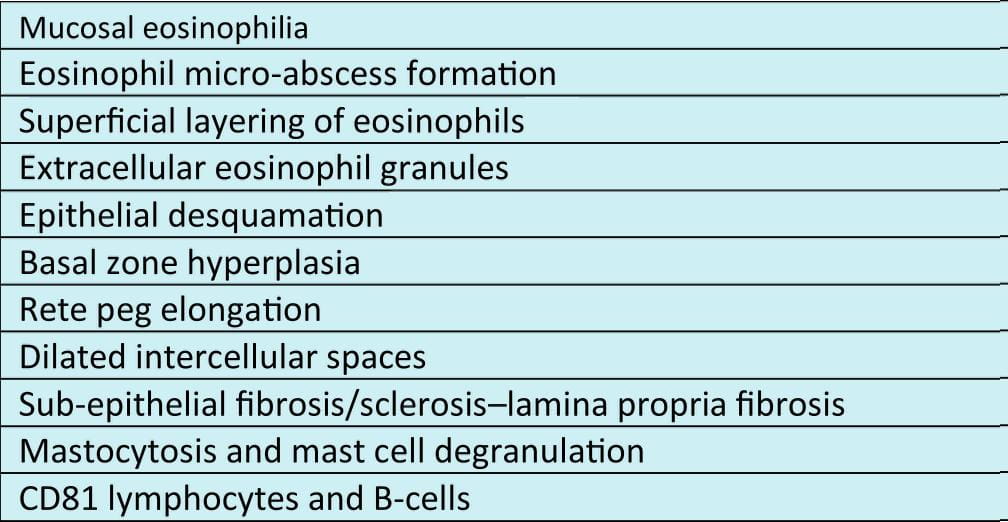
  • All histologic features should be evaluated and noted in pathology reports, including the following:
    • Peak eosinophil counts obtained from the most densely populated hpf
    • Eosinophil microabscess formation
    • Superficial layering of eosinophils
    • Extracellular eosinophil granules
    • Basal cell hyperplasia
    • Dilated intercellular spaces
    • Rete peg elongation
    • Sub-epithelial lamina propria fibrosis
  • All biopsy specimens from a single patient with EoE need not have inflammatory changes as these changes might be focal.
  • Assessment of gastric and duodenal mucosa is recommended in children as symptoms are non-specific.

Table 3: Histologic features of EoE

CPM0GHOP03888

Other Diagnostic Modalities

  • Esophageal pH monitoring is a useful diagnostic test to evaluate for GERD in patients with esophageal eosinophilia.
  • Other testing modalities do not yet offer clear clinical benefit in diagnostic testing.

Genotypic Features of Esophageal Eosinophilic

  • Clinical use of specific genotypes to predict EoE diagnosis or prognosis is not yet ready for clinical application.
  • It is, however, expected that esophageal gene expression will likely emerge as a key molecular analysis that helps differentiate EoE from other states, including GERD, to determine glucocorticoid-responding and non-responding patients and to distinguish treated EoE.

General Allergic Evaluation

  • A thorough evaluation by an allergist or immuno-logist is warranted because of the high rates of concurrent asthma, allergic rhinitis, eczema, and food allergy/anaphylaxis; the potential seasonality of EoE diagnoses; and the complex interplay among multiple allergic diatheses.

Patients with EoE:

  • 50-60% have a prior history of atopy.
  • Food hypersensitivity ranges from 15% to 43% (lgE-mediated).
  • Allergic rhinitis ranges from 40% to 75%
  • Asthma ranges from 14% to 70%
  • Eczema rangesfrom 4% to 60%

Laboratory Evaluation

Peripheral Eosinophil Counts and Eosinophil Granule Proteins

  • Increased number of circulating eosinophils (>300-350 per mm3) are observed in 40-50% patients with EoE.
  • Successful esophageal topical corticosteroid therapy can decrease peripheral eosinophilia, which correlates with tissue eosinophil numbers.

Total IGE

  • Not enough data exists to support the use of measuring total IgE levels as a surrogate marker of histologic inflammation in patients with EoE.

Aeroallergen-Specific IGE

  • A complete evaluation of patients with EoE for aeroallergen sensitization is frequently warranted in both adult and pediatric patients because this might alter clinical management.

Food-Specific IGE

  • Skin prick testing for immediate type food allergy is warranted to identify comorbid food-induced allergic disease in patients with EoE.
  • Clinical utility of IgE testing for dietary intervention in patients with EoE remains largely unknown.
  • Medically supervised food reintroduction might be necessary for patients with previous allergic reactions to a food or IgE-mediated sensitivity.
  • Patients with EoE who are found to have positive skin test results to foods should be appropriately evaluated for immediate hypersensitivity reactions.

Medical and Dietary Therapy

PPI Therapy

  • PPI therapy is useful to help eliminate GERD as a cause of esophageal eosinophilia.
  • Recommended PPI dose used to eliminate PPI-responsive esophageal eosinophilia is 20-40 mg, once or twice daily for 8-12 weeks in adults, and 1 mg/kg per dose, twice daily for 8-12 weeks in children.
  • PPIs are useful to treat patients having GERD along with EoE.

Dietary Therapy

  • All children with a diagnosis of EoE should be considered for dietary therapy.
  • The patient's lifestyle, adherence to therapy and family resources need to be considered when deciding the use of a specific dietary therapy.
  • To ensure that sufficient calories, vitamins and micronutrients are maintained, consultation with a dietitian should be encouraged.
  • Restriction of foods proved to trigger EoE might need to be discontinued indefinitely.

Corticosteroid Therapy

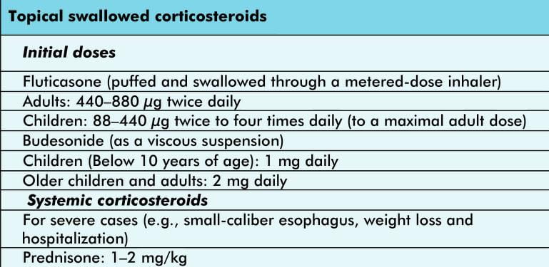
  • Topical corticosteroid therapy should be considered in all children and adults affected by EoE for both initial and maintenance therapy.
  • Disease severity, the patient's lifestyle, ability to continue the medication and family resources will determine the type and duration of steroid therapy.
  • Long-term therapy must be individualized for each patient.
  • In children, growth should be monitored carefully if they are on continual topical steroids.

Table 4: Recommended doses of corticosteroids for EoE

CPM-0GHOP0388-11

Cromolyn Solution, Leukotriene-Receptor antagonists, Biologies, and other Therapies

  • Treatment with these agents lacks clinical and histologic benefits and the side effects outweigh the benefits. Hence, these agents are not recommended.
  • Biologic agents require further clinical trials and are not currently recommended for routine use.

In EoE, prevalence of

  • Food impaction is 30-50%;
  • Stricture is11-31%; and 
  • Narrow-caliber esophagus is approximately 10%

Disease Complications

The following can point to a diagnosis of EoE

  • Food impaction
  • Unexplained esophageal strictures
  • Narrow-caliber esophagus
  • Spontaneous or endoscopic esophageal perforation

There is a risk of esophageal perforation in these patients with EoE when there is food impaction or during diagnostic endoscopy.

Esophageal Dilation

  • In selected patients, esophageal dilation with or without concomitant medical or dietary therapy can provide relief from dysphagia.
  • Trial of medical or dietary therapy is reasonable in patients without high-grade esophageal stenosis.
  • Dilation before initiation of medical therapy has been well-tolerated and effective in patients with high-grade strictures.
  • The risk of chest pain after dilation should be discussed with patients as it is significant.
  • In patients with EoE and other entities such as a Schatzki ring or peptic stricture, a more conservative approach is advised to prevent esophageal perforation.
  • Multiple dilation sessions are often required for high-grade esophageal stenosis in patients with EoE.
  • Complications of dilation can be associated with younger age and more dilations, narrowing in the upper third of the esophagus, and inability to traverse the narrowing with the scope before dilation.

Unresolved

  • Optimization of methodologies for histologic analysis of mucosal samples:
    • Number of biopsy specimens
    • Proximal versus distal biopsy specimens
    • Number of hpfs that need to be examined
    • Effect of associated histologic features on diagnostic sensitivity
    • Mean versus peak number of eosinophils
    • Size of hpf used to quantitate eosinophils
    • Use of completely filled versus partially-filled hpfs
    • Reporting eosinophil density as number per hpf versus number per unit area (mm2)
    • Eosinophil degranulation and optimal markers (MBP, EPO, and EDN)
  • Exclusion of esophageal acid/non-acid disease (PPI therapy and diagnostic testing).
  • Lack of consensus on criteria to diagnose eosinophilic disease in the remainder of the gastrointestinal tract.
  • Allergy testing in adults and children in guiding dietary exclusions.
  • Optimal endpoints of treatment (e.g., symptoms relief and histologic normalcy).
  • Frequency of endoscopy in follow-up (is it needed in asymptomatic patients?).
  • Maintenance treatment (dose and duration).
  • Validated measurements of symptoms, endoscopic findings, histology, and quality of life.

HPF: High-power fields
MBP: Major basic protein
EPO: Eosinophil peroscidase
EDN: Eosinophil-derived neurotoxin
PPI: Proton-pump inhibitor

Summary

Established

  • EoE is a clinico-pathologic disease isolated to the esophagus.
  • EoE represents a chronic, immune/antigen-mediated, esophageal disease characterized clinically by symptoms related to esophageal dysfunction and histologically by eosinophil-predominant inflammation.
  • With few exceptions, 15 eosinophils/hpf (peak value) is considered a minimum threshold for a diagnosis of EoE.
  • Endoscopy with biopsy is currently the only reliable diagnostic test for EoE.
  • An allergy evaluation is warranted in patients given a diagnosis of EoE.
  • The disease should remit with treatments of dietary exclusion, topical corticosteroids, or both.

Additional Data Required

  • Pathophysiology of PPI-responsive esophageal eosinophilia.
  • Pathognomonic features that define EoE.
  • Optimal histologic descriptive elements that characterize EoE.
  • Biomarkers and molecular signatures that aid in the diagnosis of EoE.
  • Therapeutic/predictive accuracy of skin prick and patch testing in patients with EoE.
  • Therapeutic value of dietary therapy in adults with EoE.
  • Natural history of EoE and rates and predictive indices of complications (food impaction, esophageal stricture, and esophageal narrowing).
  • Importance of treating asymptomatic patients with isolated esophageal eosinophilia.

Reference

J Allergy Clin Immunol 2011; 1 28:3-20