




If trauma is involved, establish whether it is blunt or sharp. Blunt injuries may look innocuous but may mask significant internal ocular damage. Chemical trauma should be immediately managed with thorough irrigation prior to taking any detailed history.
Acute Red Eye: Diagnosis and Treatment

4 Jun, 12


Acute Red Eye Diagnosis and Treatment
History
Trauma
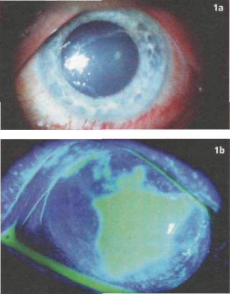
Figure 1: A corneal abrasion may be seen even without fluorescein staining. The corneal light reflex is irregular in Figure 1a. In another case (Figure 1B), an abrasion is highlighted after staining.
Small foreign bodies such as sawdust and grit, or metal foreign bodies from grinding, may blow into the eye. The precipitating event may not be obvious or offered by the patient and may need to be specifically questioned for.
Vision may vary between near-normal and severely reduced in traumatic cases. Damage may be superficial, such as a corneal abrasion (Figure 1), or intraocular haemorrhage may be present. This may be visible as a level of blood partly obscuring the lower iris - a hyphaema.
Red Eye
A red eye may be painful if inflammation such as uveitis is present, or just irritable such as with conjunctivitis, when an associated purulent discharge may also be present. A subconjunctival haemorrhage may look dramatic (Figure 2) but is often completely asymptomatic.
Figure 2: Subconjunctival haemorrhage. Note the solid red haemorrhage with distinct delineation against the white of the sclera. Resolution occurs spontaneously over 1 or 2 weeks.
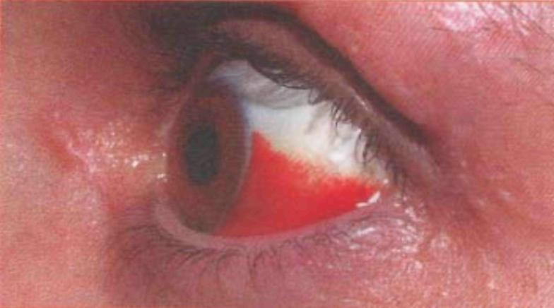
Figure 3: Acute anterior uveitis. The eye is diffusely red, particularly at the junction between clear cornea and sclera - the corneal limbus. The pupil may be irregular, and photophobia is present.
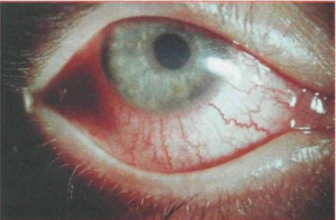
Photophobia - pain when looking at light - is typical in acute uveitis (Figure 3,overleaf ), and is usually unilateral. Bilateral red eye can be due to dry eye, and may be associated with systemic autoimmune disease such as rheumatoid arthritis. Beaware that in dysthyroid eye disease, even though the condition relates to a systemic disorder, the ocular manifestation may be unilateral.
Periorbital cellulitis in an adult, if mild,be treated with broad-spectrum oral antibiotics and reviewed the following day. If resolution is not rapid, refer, as intravenous treatment may then be required. All children require immediate urgent referral to paediatrics.
Acute glaucoma is acutely painful and the patient is often debilitated by the pain. Suspect this particularly if the patient is long-sighted (hypermetropic) and has had episodes of transient blurring or seeing haloes around light sources. If the patient's distance spectacles act as a magnifying glass, he or she is long-sighted.
Visual Symptoms
Visual symptoms are often the most challenging to diagnose.
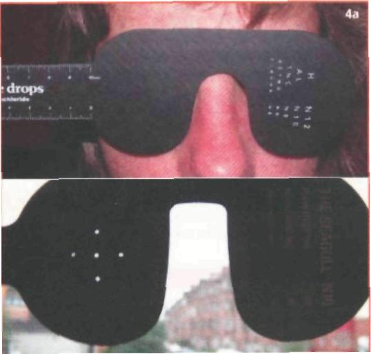
Figure 4: A pinhole substitutes for glasses. Ensure that the eye not being tested is adequately covered (Figure 4a). The patient can look through any of the holes (Figure 4b). If you do not have a pinhole, make one with a piece of paper and a few needle holes.
Reduced vision may be gradual or acute. In the elderly, cataract and macular degeneration are the most common underlying causes. A loss or distortion of central vision, particularly in the elderly, may be due to wet macular degenerative disease. Ask whether a normally straight object, such as a window or door frame, appears to have new kinks. Some of these cases can now be effectively treated if diagnosis and referral to ophthalmology is prompt.
In the elderly, the pupil may be small. Dilate it with tropicamide 1.0% to assist in viewing the fundus, particularly in patients who have diabetes. There is a fear that dilating may trigger acute glaucoma, but in reality this is extremely rare. Avoid dilation if there are features of subacute attacks such as those described above.
The appearance of new dots or cobwebs in the visual field is common. These are associated with detachment of the vitreous gel in the posterior compartment of the eye and are often accompanied by flashing light (mechanical traction on the retina). These 'floaters' may be associated with retinal holes and subsequent retinal detachment, particularly if the patient has myopia.
Transient visual loss occurs in inflammatory or embolic disease or typical migrainous fortification spectra (a pulsating serrated line moving across the visual field), often without any associated headache. Total or partial permanent loss of vision in an eye is commonly due to infarction of the retina or optic nerve. Always consider temporal arteritis, usually in those over 60, and ask the relevant questions.
Be aware that, even in apparently acute cases, questioning may reveal a significantly longer history. Establish whether the condition is new or recurrent. It is not unusual for gradual visual loss due to cataract to present as sudden loss of vision.
Having established the history, this will guide regarding examination and management.
Examination and Management
In all cases, try and document visual acuity.Test each eye individually using distance glasses if the chart used is on the wall. Ensure that the patient is not wearing reading glasses, which will make distance acuity worse.
If glasses are not available, use a pinhole (Figure 4)
Chronic intermittent, frequently unilateral irritation may result from lower lid entropion. In this condition, usually due to lid laxity in the elderly, the lid rotates inwards leading to the lower eyelashes
Figure 5: Entropion. Lower lid laxity results in the lid rotating inwards, causing the eyelashes to abrade the cornea (Figure 5a). Figure 5b demonstrates how to correct this temporarily with Steri strips or tape. Ensure the skin is dry.
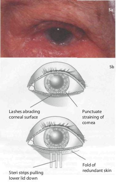
abrading the cornea (Figure 5a). An effective temporary fix is shown in Figure 5b prior to definitive surgical correction.
Chronic bilateral irritation is commonly secondary to blepharitis. The lid margins may be injected, and crusting or matting together of the lashes is seen (Figure 6). Acute exacerbations may lead to the patient presenting. Explain that the condition is long term but can be improved by methodical lid hygiene. This is best achieved by using a clean piece of cotton or lint, moistened in either saline or diluted baby shampoo. With the eyelids closed and gently pulled laterally, the lash margin
Figure 6: Blepharitis. Injected, thickened lid margins with debris on the lashes.
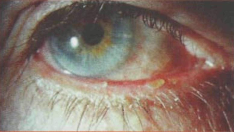
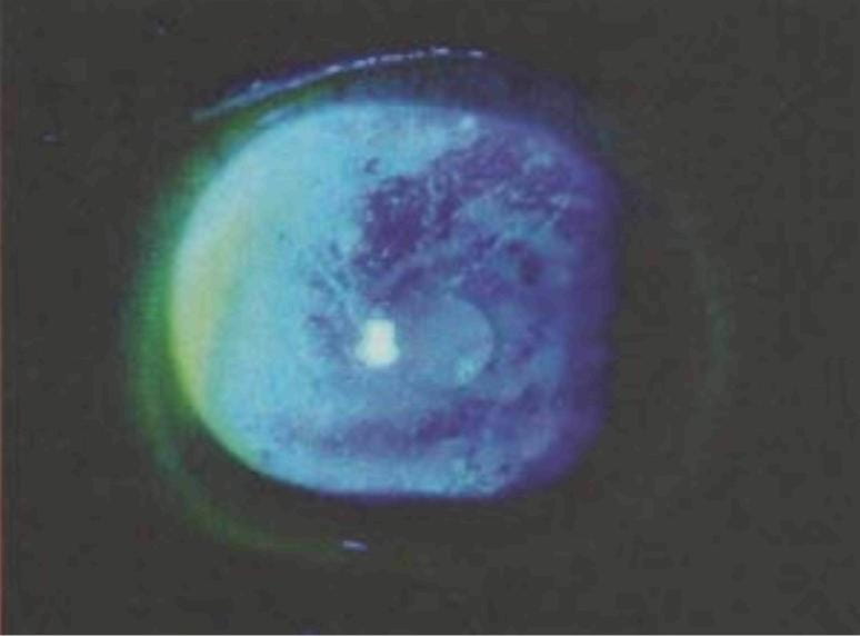
Figure 7: Typical multiple linear abrasions in the upper cornea seen with a subtarsal foreign body.
and lashes can be gently but firmly wiped,twice daily. This should be continued indefinitely. Acute exacerbations can also be treated with fusidic acid 1% ointment rubbed into the lash margin after cleaning.
A purulent discharge with conjunctivitis should be treated with chloramphenicol 0.5% drops four times daily for a week. If the conjunctivitis does not resolve or worsens with treatment, consider an allergy to the treatment used.
If a foreign body or corneal abrasion is suspected, first instil a drop of local anaesthetic such as proxymetacaine hydrochloride 0.5% or benoxinate hydrochloride 0.4%. This will enable patients to open their eyes. Ready-mixed proxymetacaine and fluorescein unims is particularly useful and fluorescein 0.25% or fluorescein can be instilled separately.
Check the cornea for an abrasion or foreign body.Figure 7 shows the characteristic multiple linear abrasions seen in the upper cornea with a subtarsal foreign body. Evert the eyelid (Figure 8), but not if you suspect a penetrating injury.
Figure 8: (a) Ask the patient to look down and keep looking down. (b) Place a cotton bud on the upper lid gently in the position shown. (c) Hold the lashes firmly and rotate them over the cotton bud. (d) Hold everything in place as you examine the eye. The lid will flick back if the patient does not continue to look down.
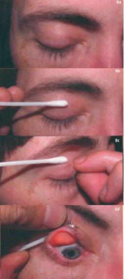
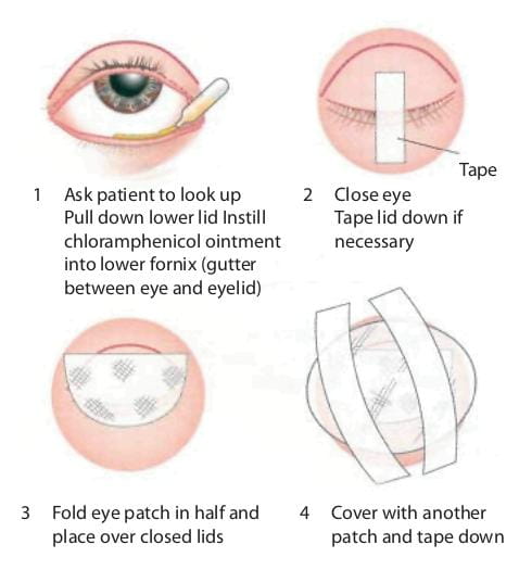
Figure 9: Technique of double patching for corneal abrasion.
Any suspected penetrating injury requires immediate referral. Be particularly suspicious of this if injury has occurred as a result of hammering or chiselling. A small penetrating injury may initially be relatively asymptomatic.
A corneal or subtarsal foreign body may be removed with a cotton bud soaked in proxymetacaine 0.5% or a similar topical anaesthetic. If more adherent, as in the case of a ferrous metallic corneal foreign body, which may have an associated rust ring, use an orange needle on a 2 ml syringe. The syringe acts as a handle.
Treatment for a corneal abrasion or following the removal of a corneal body consists of chloramphenicol 0.5% ointment and a drop of cyclopentolate hydrochloride 1.0%. Patch the eye as shown in Figure 9. Leave this on overnight. Corneal abrasions will often heal within 24-48 hours. Review if there is no improvement over 24 hours.
Suspect uveitis if light is painful (photophobia), even if the eye is not particularly red. In recurrent cases, the patient is often aware of the onset prior to any clinically obvious findings. Treatment is with topical steroids and cycloplegics such as prednisolone acetate 1% 2-hourly and cyclopentolate 1%. This should only be initiated after slit lamp examination to rule out herpetic lesions, such as a corneal dendritic ulcer. Review should be carried out within 48 hours, either in the primary care setting if a slit lamp is available, or by the ophthalmologist.
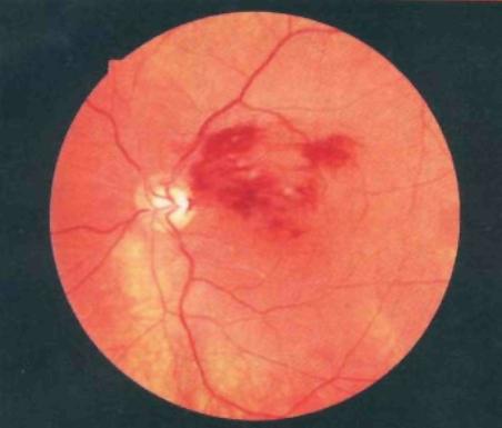
Figure 10: Branch retinal vein occlusion. The macula is involved in this case, and vision will be significantly reduced. New vessels, similar to those seen in individuals with diabetes, may subsequently develop as a result of ischaemia.
Acute glaucoma results in iris ischaemia, leading to an unreactive and semi-dilated pupil, often in association with a hazy oedematous cornea. Immediate referral to ophthalmology is required. An intravenous bolus of acetazolamide 500 mg will start the process of intraocular pressure reduction if a delay in hospitalisation is anticipated.
Visual loss due to macular degeneration can occur slowly over years, or suddenly in the case of wet degeneration, when subretinal fluid or haemorrhage affects the central visual field. The macula may show obvious features of degeneration, or these features may be subtle.The patient should be referred to the local eye unit for an urgent ocular tomogram.
Diplopia due to palsy of the lateral rectus (Vlth cranial) nerve is often associated with diabetes and hypertension. If appropriate, a course of anti-vascular endothelial growth factor injections can be initiated. This consists of a course of injections into the vitreous cavity over several weeks or months, and aims to inhibit the growth of the unstable new vessels that underlie wet degeneration.
In acute visual loss due to retina or optic nerve infarction, the eye looks unremarkable, but there may be little or no pupil response to direct stimulation. Note that, in the affected eye, the pupil may well respond briskly when the light is directed at the unaffected eye (a consensual response). An altitudinal visual field loss - superior or inferior hemi-field - is associated with retinal or optic nerve vascular occlusion. Visual loss is usually more profound in arterial occlusions, and fundoscopy may reveal a pale, infarcted retina. Hypertension and associated systemic vascular disease is common.
Aspirin 75-150 mg daily may give a degree of protection to the unaffected eye. If the event is very recent, that is within a few hours, urgent steps to reduce intraocular pressure may help, and the patient should be seen in the local eye unit as a casualty. The prognosis for visual recovery is poor.
Venous occlusions are usually evident by the presence of retinal haemorrhages (Figure 10). Vision may be only minimally affected if the macula is spared.
Hypertension, raised cholesterol and hyperviscosity states should be treated. Non-urgent review in an eye unit is required as the condition may lead to neovascular growth and complications such as glaucoma and vitreous haemorrhage.
Diplopia due to palsy of the lateral rectus (Vlth cranial) nerve is often associated with diabetes and hypertension. Horizontal diplopia occurs. Vertical diplopia and ptosis, even with a normal pupil, can occur in Illrd nerve palsy. In the elderly, a Vlth nerve palsy often resolves spontaneously within 6 weeks. If no resolution occurs, a scan to rule out a space-occupying lesion is required. A Illrd nerve palsy requires immediate referral as this can be secondary to an aneurysm.
• The history will usually pinpoint the diagnosis.
• Check the vision in each eye separately - the patient may have 20/20 vision but be blind in one eye.
• Hammering or chiselling can be associated with globe penetration despite a normal-looking eye
Reference
Webb, LA. Manual of eye emergencies, diagnosis and management (2nd edn.). Oxford, Butterworth-Heinemann, 2004.
Related Topics


