

Effect of a Modified Long-axis in-plane Ultrasound Technique in Guiding Radial Artery Cannulation in Adults


Introduction
Radial artery cannulation was traditionally performed by palpation of radial artery pulsation; conventional palpation technique (C-PT). Ultrasound-guided techniques have been widely used in various vascular cannulations because of its visual characteristics and higher success rate of cannulation compared with traditional palpation.
Aim
To evaluate the effect of the modified long-axis in-plane ultrasound technique (M-LAINUT) in guiding radial artery cannulation
Patient Profile
- Adult patients with the diameter of the radial artery were not less than 2.2mm scheduled for elective surgery were included in this trial
Methods
- Prospective, randomized and controlled clinical trial
- Conducted at Fujian Medical University Union Hospital between 2017 and 2018
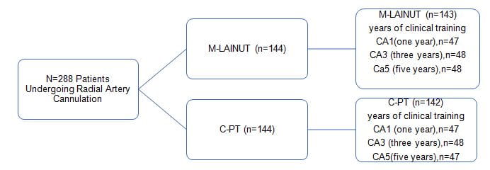
Reasons for exclusion included the reluctance to randomize (M-LAINUT, n=1), the attending anesthesiologist decided not to insert the cannula into the radial artery (C-PT, n=1), and the loss of medical records (C-PT, n=1).
- Radial artery cannulation was performed by 3 anesthesiologists with different experience.
- The primary outcome was the success of the first cannulation which was defined as the successful insertion of the cannula with 1 skin puncture for the first time and the acquisition of arterial waveforms.
- Cannula insertion failure was defined as greater than 3 cannulation attempts for a single arm or an attempted cannulation time more than 5 minutes
- Secondary outcomes included the cannulation time (seconds), the number of attempts, complications
- The number of attempts was defined as the number of skin perforations caused by the puncture needle.
Results
Table 1: Baseline characteristics
|
Characteristic |
C-PT n =142 |
M-LAINUT n =143 |
P |
|
Age, yr |
61.1 ±12.3 |
62.62 ±11.99 |
.31 |
|
Inner diameter of left artery, mm |
2.34 ±0.14 |
2.34 ±0.13 |
.65 |
|
Inner diameter of right artery, mm |
2.37 ±0.14 |
2.36 ±0.13 |
.34 |
|
Depth of Left radial artery, mm |
2.38 ±0.35 |
2.54 ±0.17 |
.27 |
|
Depth of right radial artery, mm |
2.40 ±0.36 |
2.56 ±0.30 |
.28 |
|
MAP, mm Hg |
101.7 ±18.1 |
102.80 ±18.3 |
.63 |
|
Heart rate,2 beats/min |
74.6 ±12.28 |
73.28 ±12.01 |
.36 |
|
BMI, kg/m |
22.99 ±3.33 |
22.99 ±3.28 |
1.00 |
|
Female |
82 (57.7%) |
84 (58.7%) |
.90 |
|
Left radial artery access |
95 (66.9%) |
97 (67.8%) |
.9 |
|
Medical history |
|
|
|
|
Hypertension |
17 (12.0%) |
22 (15.4%) |
.49 |
|
Coronary disease |
4 (2.81%) |
4 (2.79%) |
1.00 |
|
Diabetes mellitus |
9 (6.3%) |
9 (6.29%) |
1.00 |
|
Peripheral vascular disease |
5 (3.52%) |
6 (4.19%) |
1.00 |
|
BMI >30 kg/m2 |
6 (4.22%) |
7 (4.89%) |
1.00 |
|
Types of surgery |
|
|
|
|
Resection of lung tumor |
102 (71.83%) |
103 (72.02%) |
1.00 |
|
Urinary surgery |
22 (15.5%) |
20 (13.98%) |
.74 |
|
Others |
18 (12.67%) |
20 (13.98%) |
.86 |
Primary Outcome
- Cannulation success rates on the first attempt and total attempts in the M-LAINUT group were 91.6% and 97.9%, respectively, which were significantly higher than the 57.7% and 84.5% in the C-PT group (P<.001;odds ratio, 7.9)
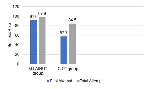
- The cannula insertion failure rate in the C-PT group was 15.5%, which was significantly higher than the 2.1% in the M-LAINUT group (P<.001)
|
Number attempts (<5 min) |
C-PT (N =142) % |
M-LAINUT (N =143) % |
P |
OR |
|
1∗ |
57.7 |
91.6 |
<.001 |
7.9 |
|
2 |
12.7 |
4.4 |
|
|
|
3 |
14.1 |
0.7 |
|
|
|
Total success rate∗ |
84.5 |
97.9 |
<.001 |
8.5 |
|
Rate of insertion failure† |
15.5 |
2.1 |
<.001 |
0.1 |
CI=confidence interval, C-PT=conventional palpation technique, M-LAINUT=modified long-axis in-plane ultrasound technique, OR=odds ratio.
∗
P-values and 95% CIs are calculated from the x2 test.
† P-values and 95% CIs are calculated from Fisher exact test.
Secondary Outcomes
- The total success rate (5minutes and 3 attempts) in the M-LAINUT group was 97.9%, compared to 84.5% in the palpation group (P<.001) (odds ratio, 8.5)
- The total cannulation time of the C-PT group was longer than that of the M-LAINUT group (Z= - 4.08, P<.001)
- The total cannulation time was shorter and the number of attempts was fewer in the M-LAINUT group than that in the C-PT group (P<.05)
- Significant reduction in the number of attempts required to cannulate the radial artery with M-LAINUT versus C-PT (mean: 1.11±0.39 vs 1.72±0.89, P<.001).
- The incidence of hematoma in the C-PT group was 19.7%, which was significantly higher than the 2.8% in the M-LAINUT group (P<.001)
- Hematoma caused by puncture was a high-risk factor for cannulation failure in both groups (P<.001; OR, 118)
Subgroup Analysis
- In subgroups (CA1, CA3, CA5), the success rate of the first attempt in the M-LAINUT group was significantly higher than that in the C-PT group
|
Subgroup |
C-PT % |
M-LAINUT % |
P |
OR |
|
CA1 |
31.9∗ |
10.63 |
<.001 |
0.06 |
|
CA3 |
33.3 † |
6.25 |
<.001 |
0.03 |
|
CA5 |
14.9 |
2.1 |
<.001 |
0.004 |
|
Total |
26.76 |
6.3 |
<.001 |
0.03 |
|
|
|
|
|
|
CA, clinical anesthetists, who are divided into 3 categories according to the year of clinical training in anesthesia after graduation from medical college: 1 year (CA1), 3 years (CA3), 5 years (CA5).
P-values and ORs (95% CIs) are calculated from Fisher exact test.
CA = clinical anesthetists, CI=confidence interval, C-PT=conventional palpation technique, M-LAINUT=modified long-axis in-plane ultrasound technique, OR=odds ratio.
∗P<.001.
† P<.001.
Conclusions
- Modified long-axis in-plane ultrasound-guided radial artery cannulation can increase the first and total radial artery cannulation success rates, reduce the number of attempts, and shorten the total cannulation time in adults
- The study demonstrates some clinical advantages for radial artery cannulation in adults, with a higher success rate of cannulation, fewer attempts and less time with modified long-axis in-plane ultrasound technique, when performed by operators with different experiences.
Reference
Medicine.2020; 99:2