

Delayed-Release Dimethyl Fumarate Favorably Impacts MRI Parameters in RRMS Patients with Sustained Benefits till 2 Years


Background
Treatment for multiple sclerosis (MS) aims at attenuating or delaying various magnetic resonance imaging (MRI) changes that mark the overall neurodegeneration and demyelination associated with the condition. The efficacy of delayed-release dimethyl fumarate (DMF) on various MRI endpoints in MS patients has been supported by various trials including DEFINE and CONFIM trials. In fact, the Phase 3 DEFINE study demonstrated a significant reduction in the mean number of new or enlarging T2-hyperintense lesions and gadolinium enhancing (Gd+) lesion activity at 2 years in patients with relapsing–remitting MS (RRMS) who were treated with twice or thrice daily DMF (240 mg).
Aim
To expand on the previously reported MRI results from the DEFINE study while focusing on additional MRI measures, delineating the time course of the effects. The study also examined the generality of this impact across a diverse patient population.
Patient Profile
- Patients (age; 18-55 years) with a diagnosis of relapsing–remitting MS (RRMS) and a baseline Expanded Disability Status Scale (EDSS) score of 0-5.0.
- Patients had at least one clinically documented relapse within 12 months before randomization, or a brain MRI scan (MRI cohort) with at least one Gd+ lesion within 6 weeks before randomization
Method
Study Design
- Double-blind randomized trial
Treatment Strategy
- 1:1:1 randomization as follows
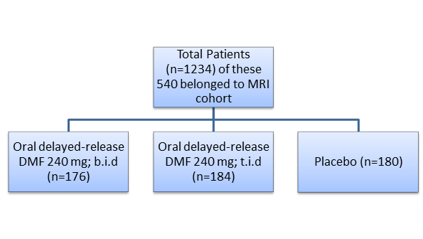
b.i.d: Twice daily
t.i.d: Thrice daily
Endpoints
Primary Endpoint
- Proportion of patients who experienced MS relapse by 2 years
Secondary MRI Endpoints
- Number of new or enlarging T2-hyperintense lesions and the number of Gd+ lesions on brain MRI.
Tertiary Endpoint
- Number of new or enlarging T2-hyperintense lesions and number of Gd+ lesions at 1 year
- Number of new T1-hypointense lesions at 1 and 2 years
- Volume of all T2-hyperintense, Gd+, T1-hypointense lesions at 1 and 2 years
- Brain atrophy at 2 years
Result
- The intention-to-treat (ITT) population comprised of 1234 patients and the MRI cohort comprised of 540 patients. Of the entire study population, 41% had received prior approved disease modifying therapy.
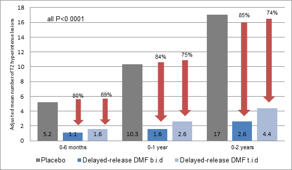
- As compared to placebo, patients in the delayed DMF b.i.d and t.i.d groups exhibited significant 80% and 69% reductions, respectively in the mean number of new or enlargingT2-hyperintense lesions right from the first MRI assessment on therapy (6 months). These significant benefits with delayed-release DMF vs. placebo were maintained at one and two years as well (all P<0.0001) (Figure 1).
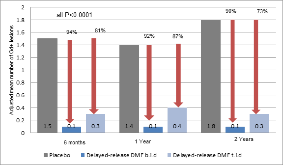
- At six months delayed DMF treatment was associated with 94% (b.i.d) and 81% (t.i.d) reduction in Gd+ lesions vs. placebo. The benefits were sustained at one and two years (all P<0.0001) (Figure 2).
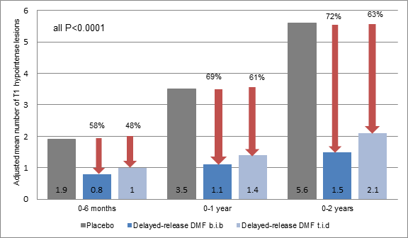
- Delayed DMF treatment was also associated with reduction in the mean number of new enhancing T1-hypointense lesions: 58 % (BID; P<0.0001) and 48 % (TID; P = 0.0005) at 6 months, by 69 % (BID; P<0.0001) and 61 % (TID; P<0.0001) at 1 year, and by 72 % (BID; P<0.0001) and 63 % (TID; P<0.0001) at 2 years (Figure3)
- Greater proportion of patients treated with delayed DMF b.i.d and t.i.d rather than placebo were free of GD+ lesions at the end of the study (93%, 86% vs. 62%).
- Patients treated with delayed DMF b.i.d and t.i.d had statistically greater reductions in mean percentage change in T2-hyperintense lesion volume from baseline to six months -3.5 and -1.7 vs. +1.6 % (BID, P = 0.0002; TID, P = 0.0035); from baseline to one year -5.8 and -3.7 vs. +6.5 % (both P<0.0001); and from baseline to two years -6.2 and -1.9 vs. +20.1 % (both P<0.0001 (Table 1).
- Delayed DMF b.i.d and t.i.d rather than placebo were also associated with a greater mean percentage change in volume (mm3) of Gd+ lesions at six months (-118.7; p<0.05, -203.2; p <0.01vs. -1.8), at one year (-88.5%, -65.9% vs. +97.4%; P<0.0001) and two years (-79.0 %, -52.6 % vs. +106.4 %; P<0.0001) (Table 1).
- Delayed DMF b.i.d and t.i.d were associated with a smaller increase in median percentage change in T1-hypointense lesion volume from baseline to six months (+1.5 and +2.5 vs. +4.3 % (both P>0.05); from baseline to one year (+5.4 and +4.7 vs. +11.6 % (BID, P = 0.0126; TID, P>0.05) and from baseline to two years (+8.4 and +12.7 vs. +26.9 % (BID, P<0.0001; TID, P = 0.0063) (Table 1).
|
Parameter |
Delayed-release DMF b.i.d |
Delayed-release DMF t.i.d |
Placebo | ||
|
% change |
P value |
% change |
P value |
% change | |
|
T2-hyperintense lesion volume | |||||
|
0-to-6 months |
-3.5 |
0.0002 |
-1.7 |
0.0035 |
+1.6 |
|
0-to-1 year |
-5.8 |
<0.0001 |
-3.7 |
<0.0001 |
+6.5 |
|
0-to-2 years |
-6.2 |
<0.0001 |
-1.9 |
<0.0001 |
+20.1 |
|
Gd+ lesion | |||||
|
0-to-6 months |
-118.7 |
<0.05 |
-203.2 |
<0.01 |
-1.8 |
|
0-to-1 year |
-88.5 |
<0.0001 |
-65.9 |
<0.0001 |
+97.4 |
|
0-to-2 years |
-79.0 |
<0.0001 |
-52.6 |
<0.0001 |
+106.4 |
|
T1 lesion hypointense lesion volume | |||||
|
0-to-6 months |
+1.5 |
>0.05 |
+2.5 |
>0.05 |
+4.3 |
|
0-to-1 year |
+5.4 |
0.0126 |
+4.7 |
>0.05 |
+11.6 |
|
0-to-2 years |
+8.4 |
<0.0001 |
+12.7 |
0.0063 |
+26.9 |
- The impact of delayed DMF on mean number of new or enlarging T2 hyperintense lesions and on the number of Gd+ lesions remained consistent across all the pre-specified population subgroups.
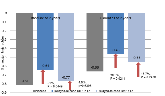
- Delayed DMF b.i.d and t.i.d attenuated the brain volume loss from baseline to two years as compared to placebo by 21 % (P = 0.0449) and 5 % (P = 0.6398). Similar attenuation of brain volume loss was also observed from 6 months to two years period with relative reductions of 30 % (P = 0.0214) and 17 % (P = 0.2478) with delayed-release DMF BID and TID, respectively.
Conclusion
- The results suggest that delayed DMF treatment reduced the inflammatory processes correlating with the relapse of MS and the burden of focal white matter disease, thus favoring the MS pathophysiology in patients with RRMS.
J Neurol. 2014; 261:1794–1802.