




Clinico-Epidemiological Pattern of Fungal Keratitis over Sixteen Years-The Indian Scenario

28 Apr, 21


Introduction
Fungal corneal ulcers are the most challenging corneal ulcers to diagnose and treat. Fungal invasion into corneal layers followed by the subsequent tissue damage is particularly devastating due to increased risk of the visual axis disruption. Clinical diagnosis of fungal keratitis can be improved by microbiological investigations, nevertheless; appropriate treatment should be initiated until the microbiological diagnosis is established. The knowledge of clinico-epidemiological pattern of fungal keratitis is thus very important.
Aim
- To analyse the results of fungal culture for patients with fungal keratitis over 16 years
- To determine the variations in the trend and distribution across ages, gender and seasons over years
Patient/ sample Profile
- Fungal culture isolates (generated from the corneal scrapings) of patients with suspected fungal keratitis attending a tertiary care teaching and research center in Northern India (n=18,898)
Methods
Study Design
- A retrospective analysis
Assessments
- A diagnosis of culture proven keratomycosis was established if:
- Same fungus was isolated in more than one inoculated media
- Fungal elements were seen in primary microscopy and fungus grew at least in one fungal culture medium
- Confluent fungal growth was obtained on the specimen inoculated in a single solid medium
Results
- Of the 18,898 suspected samples, 67.3% were for males and 32.7% for females. Of the entire study population, 21.5% (n=4069) were positive for fungal culture, of which 23.4% and 17.7% were males and females, respectively.
- Maximum number of samples (17.9%) were received for age group 41-50 years, while maximal fungal positive cultures (30.8%) were found in age group 31-40 years, followed by age group 41-50 years (25.4%), and for age above 70 years (23.2%).
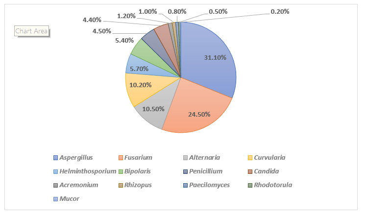
- Aspergillus species was the most common fungus (31.1%), followed by Fusarium species (24.5%), Alternaria (10.5%), Curvularia (10.2%), Helminthosporium (5.7%), Bipolaris (5.4%), Penicillium (4.5%), Candida (4.4%), Acremonium (1.2%), Rhizopus (1.0%), Paecilomyces (0.8%), Rhodotorula (0.5%) and Mucor (0.2%) (Fig. 1).
Fig. 1: Spectra of fungal species isolated from the culture
- Fungal culture positivity and relative frequency of fungi remained nearly stable over the study duration, except for Rhodotorula species, which showed a rise 2014 onwards.
- The numbers of culture proven fungal keratitis cases were high from August to November (Monsoon), with the number being highest in September and October.
- The culture positivity was significantly higher in the colder months (December to February) than hot and dry months (May and June)
Conclusions
- This Indian study is the largest compilation of epidemiological and microbiological features of fungal keratitis, that provides valuable insights for improving the management of mycotic keratitis.
- Understanding the pattern of variations of various fungal species across different age groups, and in different regions and seasons may improve the management and thus the visual outcomes in the patients with mycotic keratitis.
J Infect Public Health. May-Jun 2019;12(3):367-371.
Related Topics
More From Area Of Interest
You may also like

13 May, 26

17 Apr, 26
Our Research
23 Feb, 26

23 Feb, 26
11 Feb, 26
11 Feb, 26
11 Feb, 26
Latest Items
Journal Scans
23 Jul, 26

23 Jul, 26

23 Jul, 26

23 Jul, 26


