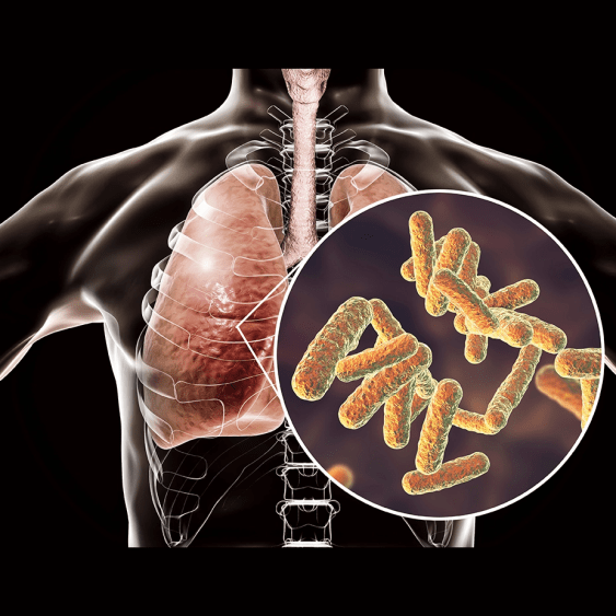
This session provides a comprehensive overview of the Consequences of Zika Infection in the Brain. The Zika virus is a flavivirus spread by mosquitoes. The first three open reading frames make up the virus particle, which has a membrane-bound monomer dimer and a hexamer in solution. The NS1 protein is crucial to the disease tropism or target of the virus, especially in the brain and placenta vasculature. The Zika virus emerged from a forest or jungle sylvatic cycle to a human mosquito-human cycle. It causes similar acute clinical pictures as other mosquito-borne viruses, especially fever, macular malar rash, headache, and myalgia. Zika also has conjunctivitis as one acute phenomenon, but not always. The virus is transmitted sexually, perinatally, through blood and blood donations. The strains have two lineages of the virus, Asian and African, and it emerged in South America through a tight bottleneck. Zika is associated with Guillain-Barre syndrome and congenital Zika syndrome. The congenital Zika syndrome is associated with microcephaly and other symptoms, such as calcifications, Ventriculomegaly, ocular abnormalities, congenital contractors, and general growth restriction. Pregnant woman who travelled to South America was found to have high levels of Zika virus, and ultimately terminated her pregnancy due to fetal abnormalities. The various effects of Zika virus on fetal development, including abnormal language and motor skills, with males being more affected than females. The virus can infect the placental trophoblasts and hoffbauer cells, which disrupts the placenta endothelial barrier, leading to the transmission of the virus to the fetal brain. The virus inhibits interferon signalling. It targets human STAT2 through the NS5 protein, which leads to fetal brain damage in humans and primates. The virus disturbs the division plane and disrupts the ventricle zone, resulting in cortical thinning. It prefers the brain and progenitor cells, causing microcephaly in 5% of infected mothers. A study in Brazil found that non-identical twins with the same environmental exposure had different outcomes, with one developing microcephaly and the other developing normally. They found that infected cells had reduced cell growth and deregulated pathways. Genetic factors may play a role in predisposing individuals to the virus, although this is not entirely clear. Immunity to the virus can be both IgG and IgM. In addition, there is a possibility of making a monoclonal antibody that is protective against the virus. According to research, the presence of specific amino acids in the American Asian strain of the Zika virus may make it more pathogenic than the African strain. However, this conclusion was based on experiments conducted on a strain that had been artificially altered through multiple passages, and may not reflect the natural African strain accurately. More recent studies have suggested that the African strain could cause more fetal loss but fewer congenital disabilities, perhaps because it is more virulent. It is also believed that the African strain may be less prone to the Zika virus, which could impact African populations differently. Overall, the session highlighted the different impacts of the Zika virus on populations, such as GBS and CZS, with microcephaly occurring in 4% of infected pregnant mothers. In addition, prolonged infection in placental cells can cause fetal brain infection, and African strains of the virus may cause more fetal loss due to their higher virulence. The virus can also affect the CNS by causing neural apoptosis and disrupting neural progenitor differentiation, and the likelihood of congenital disabilities is influenced by the viral lineage.
Immune-mediated Control of Alphavirus Replication in the CNS
Alphaviruses, from a pathogenic perspective, can be broadly categorized into two types. The ones circulating in the Americas, such as Western, Eastern, and Venezuela encephalomyelitis, affects the nervous system. On the other hand, the viruses found primarily in Europe, Africa, and Asia are more likely to cause rash and arthritis, with fewer complications in the nervous system. The Alphavirus genome is similar in length to flaviviruses at approximately 12,000 nucleotides. It produces non-structural proteins from genomic RNA and generates a distinct RNA. Sindbis virus, a type of Alphavirus, is often studied due to its neurotropic nature and ability to replicate in neuron cytoplasm. This allows for the investigation of viral clearance during recovery from encephalomyelitis. Sindbis virus infection in mice leads to recovery without neurological abnormalities, resulting in a long-lived population of vital cells. The replication cycle of Alphavirus typically involves the virus entering the cell through an acidic endocytosis pathway. Inside the cell, the virus synthesizes viral RNA and non-structural proteins and produces subgenomic RNA for structural glycoproteins and buds from the infected cell's plasma membrane. Researchers transferred T-cells and serum from recovered mice with a genetic mutation preventing immune response to study viral infection resolution. Serum-containing antibodies cleared the virus from the nervous system, while T-cells were ineffective. Low levels of viral RNA persisted in mice's lifetime. Further analysis revealed that the antibodies targeted a glycoprotein called E2 on the virus surface. Past studies demonstrated that antibodies targeting persistently infected neurons act non-catalytically without harming neurons and require bivalent antibodies for efficacy. These antibodies were found to restore host protein synthesis, reverse Alphavirus infection effects on Na+K+ATPase, and restore cell responsiveness to interferon-alpha. This antibody can bind to infected cells, signal to alter the intracellular environment in neurons, inhibit Alphavirus replication, and block virus exit. The reverse phase protein array (RPPA) was used to study signaling changes in SINV-infected neurons after adding an anti-E2 antibody. The results showed transient NF kappa B pathway activation within 30 minutes, followed by its shutdown. Prolonged phosphorylation of STAT3 on tyrosine was also observed, as confirmed by Western blot. Based on the RPPA data, the proposed model suggests that an IL-6 family cytokine may be involved in this process, as these cytokines are known to be activated by NF kappa B and result in JAK-STAT3 phosphorylation. Leukemia Inhibitory Factor (LIF), a protein in the IL-6 cytokine family, is expressed in the nervous system by neurons and glial cells. It activates STAT3 phosphorylation through its receptor and has various roles, including promoting neurogenesis, neuronal survival, and suppressing inflammation. In an experiment, the effects of LIF on virus replication were tested, and it was found that when LIF was added alone, it had no impact. Combined with an antibody, it amplified its effect, indicating synergy in inhibiting virus replication. A new model has been developed involving the activation of NF kappa B and the production of LIF, along with antibodies and signaling for JAK-STAT-3 phosphorylation to inhibit virus replication. Viral RNA is labeled in green to track activity, and the addition of antibodies reduces viral RNA and promotes cell survival. However, some cells still show green color, indicating persistent viral infection, as these cells no longer express E2 on their surface, evading the antibody response. In mice infected with a virus, the virus is cleared within eight days by anti-E2 antibodies and interferon-gamma, but viral RNA persists in the nervous system. Inflammatory responses involving CD8 T-cells, CD4 T-cells, and B-cells are observed during viral clearance, and these immune cells persist in the same areas where the virus was present. This immune cell accumulation occurs within the parenchyma, indicating ongoing immune response even after viral clearance. During early viral infection, B cells produce IgM antibodies which are important for clearing the virus. IgM in cerebral spinal fluid can predict recovery or negative outcomes. After a few days, B cells also produce IgG and IgM antibodies. Some B cells persist in the immune system for months, producing IgG and IgA antibodies. These long-term B cells can be found in cerebral spinal fluid even after recovery. Currently, it is understood that viruses can infect neurons, but viral replication and antibody production usually cease within a week or two. However, viral RNA and immune cells may persist in the nervous system, possibly preventing virus reactivation post-recovery. In summary, anti-E2 antibodies trigger NF kappa B signalling, which synergizes with lift receptor and E2 signalling to activate STAT3, leading to decreased viral RNA structural protein production and inhibition of virus replication. Changes in RNA production are observed, including persistent RNA in B-cells producing antibodies and T-resident memory cells producing interferon-gamma.
European Congress of Clinical Microbiology and Infectious Disease 2023, 15th April - 18th April 2023, Copenhagen, Denmark