Surgical Insight: Part 2 - Keratoconus and its Management


Introduction
Surgical Insight Issue 5 had provided a brief on keratoconus, its etiology, pathogenesis, risk factors, diagnosis, imaging and an introduction to its management. The present issue continues on the management of keratoconus highlighting the surgical procedures used for the management of keratoconus.
Surgical Management Procedures
Corneal Cross-linking
Corneal collagen cross-linking (CXL) has emerged as a promising treatment to slow or even stop the progression of keratoconus and other ectatic diseases such as pellucid marginal degeneration or iatrogenic keratectasia after corneal refractive surgery.1
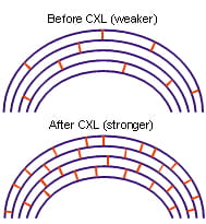
The standard protocol for collagen cross-linking (CXL) includes epithelial abrasion, the administration of a photosensitizer, riboflavin (vitamin B2, 402.7 mOsm/L) for 30 min combined with ultraviolet A (UVA) irradiation (3 mW/cm ; 5.4 J/cm ; and 370 nm) for 30 min, while riboflavin instillation continues. This technique induces free radicals which catalyze a reaction resulting in additional covalent bonds between collagen molecules with subsequent biomechanical stiffening of the cornea of over 300%.1
It has been recommended not to perform this technique in corneas thinner than 400µm as toxic reactions could take place in the corneal endothelium.2
Clinical Indications
Useful in progressive keratoconus indicated as increase in Kmax at the apex of keratoconus of 1 diopter (D) in 1 year, deterioration of visual acuity, or the need for new contact lens fittings more than once in 2 years. In US studies, CXL was performed when an increase of ≥1 D in the steepest K-reading and/or the manifest cylinder, or an increase of ≥0.5 D in refraction spherical equivalent was evident over 24 months.1
Patient Selection
Patients with worse preoperative corrected distance visual acuity (CDVA) and higher K-values, particularly with a CDVA of 20/40 or worse or a maximum K of 55.0 D or more, were most likely to have improvement after CXL.1 Age older than 35 years and a preoperative CDVA > 0/25 were identified as significant risk factors for complications. A high preoperative maximum keratometry reading >58.0 D was a significant risk factor for CXL failure. Moreover, pregnant and lactating women are typically excluded from treatment. In pediatric patients, keratoconus may present more aggressive with high rates of progression to keratoplasty. CXL should be considered earlier in these young patients.1
Long-term Clinical Studies
Several long-term studies on subjects who underwent corneal cross-linking have reported an improvement in best corrected visual acuity, a flattening of keratometric readings and a significant reduction in cone progression. Also, this technique has been successfully used in combination with other surgery techniques, such as corneal rings segments. The use of corneal cross-linking, however, has been associated with a decrease in the number of keratocytes immediately after treatment, followed by a progressive recovery post-operatively reaching baseline levels six months after treatment, accompanied by an increase in the density of stromal fibres.2
Complications
Complications in CXL treatment are reported to occur in 2.9-3.5% of patients. They include loss of BCVA, sterile infiltrates, stromal melting and scarring, bacterial keratitis, and reactivation of herpes simplex virus (HSV)-keratitis. The failure rate of CXL (percentage of eyes with continued progression) was 7.6% in a study by Koller, et al.1
Adverse Effects
Reported adverse effects with collagen crosslinking include bacterial, fungal, acanthamoeba, and sterile keratitis. Kymionis et al reported significant endothelial cell loss after crosslinking in thin corneas. Spoerl et al have described safety-related guidelines to be followed during the crosslinking procedure. These include3:
- Epithelial removal
- Application of 0.1% riboflavin solution for 30 minutes before ultraviolet exposure
- A homogenous ultraviolet irradiance of 3 mw/cm2 with a wavelength of 370 nm
- Minimal corneal stromal thickness of 400 μm.
Damage to the endothelium, lens, or retina is not expected if these criteria are fulfilled. Of great concern are reports of persistent corneal edema and corneal decompensation, along with signs of damage to anterior segment structures following crosslinking in concordance with established protocols and guidelines.3
Safe clinical application of CXL must adhere to the following requirements4
- To facilitate diffusion of riboflavin throughout the corneal stroma, the epithelium should be removed or a sufficient permeability of the epithelium must be guaranteed
- 0.1% riboflavin solution should be applied for at least 20 min before UV exposure (during UV exposure, the riboflavin serves as both a photosensitizer and a UV blocker)
- The UV dosage of 5.4 J/cm2 with a wavelength of 370 nm must be homogenous
- The cornea to be crosslinked must have a minimal thickness of 400 mm to protect the endothelium
Protocol Modifications
Modifications to the original Dresden protocol include use of hypo-osmolar riboflavin to swell thin corneas artificially, transepithelial crosslinking using different compounds designed to improve riboflavin penetration or using iontophoresis, and attempts to reduce overall duration of the procedure by increasing ultraviolet radiance. The safety and efficacy of these modifications is unproven.3
Transepithelial CXL (TE CXL)
TE CXL has been propagated to avoid epithelial debridement, but in vitro studies showed only low concentrations of riboflavin into the corneal stroma without epithelial removal. Functional results after TE CXL showed keratoconus instability and regression of the primarily obtained result. TE CXL was less effective than standard CXL when using proparacaine drops 0.5% preserved with benzalkonium chloride (BAC) 0.005%. Published evidence suggests using riboflavin combined with ethylenediaminetetraacetic acid 0.01% and trometamol to enhance epithelial penetration.1
TE-CXL may be reserved for pediatric cases, uncooperative patients, and thin corneas.1
CXL in thin corneas
Thin corneas with a stromal thickness of less than 400µm are excluded from the standard treatment so as to
avoid damaging the endothelial cells with the UV radiation. Because many patients with keratoconus have a corneal
thickness of less than 400 µm, various methods of CXL have been developed to treat this patient population
with varying degrees of success. Methods are described below.4
CXL with Hypoo-smolar Riboflavin
Corneal thickness can be increased to 400 µm by applying a hypo-osmolar riboflavin solution to produce swelling without reaching the toxicity threshold of the endothelium. The CXL effect is comparable to that in a cornea with a thickness of 400 µm because the anterior stroma barely swells while the posterior stroma swells greatly. Using this technique, it is possible to treat corneas with a minimum stromal thickness of 320 µm. Positive clinical results from this technique have been recorded.4
CXL with Reduced Irradiation Time
It has been suggested that the irradiation dose can be decreased in accordance with the thickness of the stroma
(shorter irradiation time at an irradiation intensity of 3 mW/cm2) so as not to exceed the toxicity threshold of
0.63 J/cm2 for the endothelium. This method has been used primarily for the CXL of thin corneas when no hypo-osmolar
riboflavin solution was available for topical application.4
- To protect the endothelium from very high irradiation intensity, the epithelium is not removed from the thinnest place on a surface with a corneal thickness of <400 µm. However, with the “customized epithelial debridement technique,” not enough riboflavin penetrates below the epithelium; thus, no keratocyte changes and no clear line of demarcation (as is present with complete epithelium removal) could be found, which suggests an insufficient biomechanical effect.4
- With a brief application of riboflavin to the surface, a sufficient concentration in the anterior stroma should be achieved without riboflavin reaching the endothelium. Because the toxicity threshold of the endothelial cells is much higher without riboflavin, the endothelial cells would thus be protected, even in thin corneas. However, a prerequisite for this procedure is a short irradiation time with a high irradiation intensity (equal dose) to keep the diffusion time short. 4
- An increase in the concentration of riboflavin to 0.2% leads to a greater absorption of UV light in the anterior stroma and a decrease in UV exposure of the endothelium.
- Combinations of various techniques described above, for example 1 + 5 or 3 + 5, increase safety during the CXL of thin corneas.4
CXL with Improved Intensity Profile
The intended depth of CXL using current light sources is achieved only within the central area of the cornea. To provide CXL to the peripheral cornea, the UV beam should either have an improved intensity profile or may have to be decentered. New CXL devices with an optimized beam profile to increase the CXL depth in the periphery, and thus the volume which is cross-linked, are now available and are evaluated in clinical trials.1
Combination with Other Procedures
Combinations of CXL with implantation of intrastromal corneal ring segments ± transepithelial phototherapeutic keratectomy, has been reported in small case series with promising results. It seems that intrastromal corneal ring segments (ICRS) implantation followed by CXL results in greater improvement of keratoconus than CXL followed by ICRS placement. Moreover, laser power must be modified following CXL suggesting channel dissection and ICRS implantation before or concurrent with CXL. A clinical trial comparing CXL alone to CXL combined with Intacs in patients with keratoconus and post-LASIK ectasia is currently underway in the USA.1
Keratoplasty
Corneal transplantation has evolved rapidly over the past decade with an increase in anterior and posterior lamellar keratoplasty replacing penetrating keratoplasty. Zirm performed the first successful penetrating keratoplasty in 1905, and the subsequent 90 years witnessed advancements in sutures, antibiotics, corticosteroids, operating microscopes, and eye-banking to improve surgical outcomes. Penetrating keratoplasty remains the most common tissue transplantation performed worldwide. Primary indications for surgery include keratoconus, repeat grafts, aphakic and pseudophakic bullous keratopathy, dystrophies, infections, and trauma.5
In recent years, Descemet membrane endothelial keratoplasty (DMEK) has gained interest as it eliminates the corneal stromal interface, which may limit visual acuity after Descemet stripping automated endothelial keratoplasty (DSEK). Endothelial keratoplasty—DSEK or DMEK—was created to treat endothelial disease, while deep anterior lamellar keratoplasty (DALK) was developed to target stromal disease.5
Figure 2: Schematic overview displaying (A) a virgin cornea and (B–E) different keratoplasty procedures: (B) PK, (C) DALK, (D) DSAEK, and (E) DMEK

Indications for Keratoplasty in Keratoconus
Poor visual acuity with contact lenses, contact lens intolerance or inability to fit/wear contact lenses, and nonresolving corneal hydrops,3 corneal scarring, corneal keratometry steeper than 55 D, corneal astigmatism > 10D, early stage of keratoconus development.2
A large, prospective, multicenter, Collaborative Longitudinal Evaluation of Keratoconus study reported a 12% rate of keratoplasty over an 8-year follow-up period.3
Penetrating Keratoplasty
For quite a few decades, penetrating keratoplasty has been successfully used for visual rehabilitation in keratoconus. Reasonably good visual and refractive outcomes with low complication rates have been consistently reported. Keratoconus is amongst the best indications for doing a penetrating keratoplasty, with long-term graft survival rates surpassing those for any other indication. The graft survival rates at 1, 5, 10, 15, and 20 years, respectively, are 97%, 95%, 89%, 77% and 46%.3
Deep Anterior Lamellar Keratoplasty (DLK)
In recent years, DALK has emerged as an attractive alternative to penetrating keratoplasty for keratoconus. Unlike penetrating keratoplasty, DALK is not a full-thickness corneal replacement procedure.
- Technique: The epithelium and stroma of the host cornea, preferably up to the descemet membrane (DM), are removed, thereby preserving native endothelium. This can be achieved either by manual dissection or by a “big bubble” technique, which uses an air bubble to dissect the plane between corneal stroma and the DM. The donor cornea with the DM stripped off is then sutured on like a penetrating keratoplasty.
- Advantages: DALK has the advantages of essentially being an extraocular procedure and retaining the host endothelium, thereby obviating the risk of endothelial graft rejection and probably improving graft survival. 3
- The surgery is technically challenging to perform, has a significant learning curve, and demands greater operating time compared with penetrating keratoplasty.
- Complications: intraoperative DM perforation, which may necessitate conversion to penetrating keratoplasty, postoperative DM detachment, and interface haze. Also, baring of the DM may not be possible in cases with deep stromal scarring or prior hydrops, resulting in suboptimal visual outcomes due to retained stroma. Nonetheless, pre-descemetic DALK may be a good alternative to penetrating keratoplasty in such cases.3
- Surgical Outcomes: outcomes are similar to penetrating keratoplasty in terms of visual acuity and astigmatism, with about 80% of patients achieving a best-corrected visual acuity of 20/40 or greater in most series. DALK is equivalent to penetrating keratoplasty in terms of refractive error, and is superior for preservation of endothelial cell density.3
- Recommendation: DALK should be performed as the standard of care surgery when keratoplasty is required in keratoconus.3
Femtosecond Laser-Assisted Corneal Transplantation
- Newer technology with the femtosecond laser is being used for reproducible, customized transplant designs in the dissection of both donor and recipient corneas.
- Its use in anterior lamellar keratoplasty and penetrating keratoplasty may improve wound stability and reduce postoperative astigmatism due to increased accuracy and predictability.
- Reproducibility of lamellar cuts may decrease the need for corneal sutures, leading to earlier visual rehabilitation and less suture-related complications.
- Whereas the potential for better optical results is encouraging, current use is limited by high costs and lack of portable equipment.5
- Surgical Outcomes in Keratoconus:
- In 2014, a non-comparative case series study results indicated that femtosecond laser-assisted DALK could improve UCVA and BCVA in patients with anterior corneal pathology. This approach showed promise as a safe and effective surgical choice in the treatment of keratoconus and post-LASIK keratectasia.6
- Another study showed that Femtosecond laser assisted intrastromal corneal ring segment implantation was a safe and effective method for the correction of keratoconus7
Descemet Stripping Automated Endothelial Keratoplasty (DSAEK)
- DSAEK has now become a popular alternative to penetrating keratoplasty and is the procedure of choice for endothelial dysfunction.5
- Advantages Over PK: less ocular surface complications and structural weaknesses, minimizing wound dehiscence and induced astigmatism, allows faster visual rehabilitation and avoids suture-related complications.5
- The primary complication after DSAEK is donor detachment and dislocation. Dislocation rates vary by surgeon experience and technique with rates ranging from 4 to 50%. Techniques such as prolonged full-chamber air bubble, stab incisions, surface sweeping to reduce interface fluid, peripheral scraping, and longer postoperative supine positioning have been suggested to decrease dislocation rates.5
- Visual Outcomes: Visual acuity outcomes are good with minimal astigmatism induced, although there can be a postoperative hyperopic shift up to 1 diopter. Most patients achieve corrected distance visual acuity of 20/40 or better after DSAEK, but not many achieve 20/20 visual acuity. The suboptimal vision may be attributable to aberrations from the stromal interface that can degrade vision.5
Photorefractive Keratectomy (PRK)
- Photorefractive Keratectomy has the benefit of Leaving a thicker residual stromal bed after surgery than LASIK and has generally been considered to be a safer option in suspect or thin corneas.8
- Cennamo and colleagues reported on the treatment of mild to moderate keratoconus with PRK with topographically supported customised ablation. Post-operatively the mean uncorrected vision and visual acuity were significantly increased and the mean keratoconic topographic indices were all significantly lower. There were no complications and the results were stable at 24 months, although the authors conceded that longer follow-up was required.8
- Studies have reported good outcomes of photorefractive keratectomy in keratoconus suspect eyes, with excellent uncorrected visual acuity and predictable refractive outcome in a majority of eyes. None of the studies report progression of ectasia after the procedure. Results need to be interpreted with caution, because not all included cases seem to be representative of keratoconus. It is suggested that topography-guided ablation may be the best choice for such cases. Topography-guided conductive keratoplasty has been proposed as a means of reshaping the cornea in advanced cases of keratoconus.3
- LASIK should not be considered for patients with forme fruste or keratoconus, as well as patients with a family history of keratoconus.5
Additive Procedures
Alternative approaches to the ablative procedures of the excimer laser are the additive procedures, which include Intra-corneal rings segments (ICRS) and intraocular lenses (IOL). As these procedures do not involve the removal of corneal tissue, there is less risk of weakening the structural integrity of the cornea and triggering progression of ectasia, as well as offering some degree of potential reversibility of the procedure should there be any complications.8
Intra-Corneal Rings Segments (ICRS)
- Uses: This technique is mainly used to treat mild to moderate keratoconus to improve contact lens tolerance.
- Intra-corneal ring segments are crescents shaped half rings made of polymethyl methacrylate (PMMA) that are inserted into channels deep in the corneal stroma. The goal of insertion of intra-corneal ring segments is not to eliminate corneal disease but to delay or eliminate the need for a corneal transplant by decreasing corneal abnormality and restoring contact lens tolerance. They may also have a use for correction of low myopia in forme fruste keratoconus.8
- Prerequisites: non-progressive keratoconus, an absence of significant central corneal scarring, and a minimum pachymetry of 450 μm at the site of implantation.2,3
- Technique: It consist of implantation of one or two polymethyl methacrylate segments in the corneal stroma to achieve central flattening of the surface, using channels created mechanically or by means of a femtosecond laser. 2,3
- Commonly Used Types of ICRS: Intacs, Kerarings and Ferrara rings. When comparing Intacs, Kerarings and Ferrara rings, no one type is overall significantly better than any other.8
- Outcomes: This surgical option has been associated with an improvement in uncorrected and best corrected visual acuity and a decrease in high order corneal aberrations, especially coma.2 Quality of vision may not be improved much in early cases or in those with a good contact lens fit, and their utility in advanced keratoconus cases is doubtful.3
- Angle closure disease and diseased corneal endothelium are absolute contraindications to the use of ICLs.2
- Complications: epithelial defects, anterior and posterior perforation, asymmetric segment placement and extension of the incision toward the central visual axis or the limbus6, erosion, infection, corneal haze, neovascularization, melting, and loss of visual acuity.3
Phakic Intraocular Lenses
- Alternatives to intra-corneal implants are intraocular lenses. They offer correction of higher spherical and astigmatic errors than intra-corneal ring segments and have a much faster rehabilitation than corneal transplantation.8
- Disadvantages: risk of endothelial damage and difficulties with intraocular lens power calculations due to low repeatability of keratometric measurements, less suitable for eyes with advanced keratoconus or highly irregular astigmatism.8
- Anterior Chamber Phakic Intraocular Lenses: includes iris-supported lenses like the Artisan and Verisyse phakic intraocular lenses. Standard (Artisan, Verisyse), toric (Artisan Toric), foldable (Artiflex) and foldable toric (Artiflex Toric) versions are available. 8
- Large amounts of spherical error (up to -22.00 D) can successfully be corrected, as well as highly significant improvement in uncorrected vision. Many patients also achieve increased visual acuity. Cylindrical correction is good at lower levels of astigmatism
- Complication rates are low but include halos and glare in low light, endothelial cell loss (up to 10 to 11 per cent at up to five years after intraocular lens insertion), cataract formation, dislocation of the intraocular lens and pigment dispersion
- Toric intraocular lenses ideally should only be considered in patients with mild keratoconus and low levels of relatively regular astigmatism.5
- Posterior Chamber Phakic Intraocular Lenses are placed between the iris and crystalline lens. Options include the Visian Implantable Collamer Lens (ICL) and the Visian Toric Collamer ICL (TICL).8
- visual and refractive results are excellent, with studies showing 96 to 100 per cent of eyes achieving ±1.00 D of the intended spherical target, post-operative residual cylinder less than 1.00 D in 87 per cent and significant improvements in uncorrected vision and visual acuity
- Potential complications include cataract in up to 33% of eyes, pigment dispersion syndrome, pupil block glaucoma, chronic uveitis and iris ovalisation
A disadvantage with any type of phakic intraocular lens is that, unlike corneal-based treatments that aim to normalise the shape of the keratoconic cornea, the intraocular lens can correct only spherical and cylindrical error. It is known that eyes with keratoconus have significant aberrations that affect visual quality, with higher levels of vertical coma, primary coma and coma-like aberrations in keratoconic eyes than in normal eyes.
The implantation of an intraocular lens for the management of keratoconus is normally undertaken in combination with other types of corneal refractive surgery techniques, such as corneal rings or keratoplasty, as intraocular lens implantation does not normally affect corneal shape and cone progression. Furthermore, the combination of these techniques, which allows the correction of high levels of astigmatism by placing an intraocular lens in the anterior or posterior chamber, has been used with relative success in a limited number of subjects, normally intolerant contact lens wearers, who has shown significant improvement in visual acuity.2
Refractive Lens Exchange8
- Once patients have reached the age of presbyopia or in patients with detectable cataract, lens removal with insertion of an intraocular lens is a better option than a phakic intraocular lens.
- Visually significant cataract has been observed to occur in patients with keratoconus at a younger age (mean 55.3 years) than in the general population (mean 69.6 years).
- Toric Intraocular Lenses Available: includes ‘off the shelf’ lenses such as the Acrysof Toric intraocular lenses with cylindrical powers up to 6.00 D and spherical powers from +6.00 to +30.00 D and more customised lenses, such as the Rayner toric intraocular lenses with cylindrical powers up to 11.00 D and spherical powers from -10 to +35 D. This enables correction of the high refractive errors associated with keratoconus.
- Intraocular Lens Power Calculation: The best lens formula to use in keratoconic eyes has yet to be established. A study comparing three formulae (SRK, SRKII and SRKT) in keratoconic eyes found that in mild keratoconus the most accurate intraocular lens power was found by using the SRKII formula. In moderate to severe cases no valid conclusion could be made.
- If the pre-operative difference between RGP contact lens corrected vision and spectacle corrected vision is high, a toric intraocular lens may not be suitable.
- Surgical Outcomes: Cataract extraction with implantation of these toric intraocular lenses has been reported to have good results, with significant improvement of uncorrected vision and spherical and cylindrical refractive error, although it has also been noted that patients with moderate to severe keratoconus often still require rigid gas-permeable (RGP) lenses after surgery.
Combination of Procedures
In recent times, a spate of combinations of procedures, such as collagen crosslinking, intracorneal rings, ICLs, and photorefractive keratectomy have been reported.
Combining corneal collagen cross-linking with intraocular lens insertion8
- One of the key issues for any intraocular lens procedure is the stability of keratoconus post-operatively. Combining corneal collagen cross-linking with intraocular lens insertion may offer the possibility of improved outcomes. In general, it is believed that the best outcomes are achieved when collagen cross-linking is performed sometime prior to intraocular lens implantation, particularly as corneal collagen cross-linking produces statistically significant reductions in keratometric values and refractive errors over time
- If collagen cross-linking has not been done and keratoconus is seen to progress after intraocular lens insertion, then the collagen cross-linking can be safely done in the presence of an intraocular lens.
Combining Intra-Corneal Ring Segments with Intraocular Lenses8
- This offers the possibility of improved outcomes by combining the effects of improving corneal shape with the intra-corneal ring segments with the ability of the intraocular lens to correct high spherical and cylindrical refractive error. The insertion of the intra-corneal ring segments can be done simultaneously with the intraocular lens or at a date earlier to the intraocular lens insertion.
- Benefits of Simultaneous Surgery: better view for intraocular lens insertion before the rings are inserted.
- Benefits of Sequential Surgery: the option to allow the patient to undergo a contact lens trial after insertion of intra-corneal ring segments before committing to additional intraocular lens insertion and keratometric readings can be taken after insertion of the ring segments, which theoretically allows for better prediction of the intraocular lens power before implantation.
Combination of implantation of intracorneal ring segments followed by sequential same-day PRK/collagen cross-linking
- It might be a good option for improving visual acuity in select patients with keratoconus8
Maximum interest seems to be focused on combining collagen crosslinking with photorefractive keratectomy. Same day collagen crosslinking combined with topography-guided photorefractive keratectomy has been found to be superior to sequential procedures for visual rehabilitation. Other approaches tried for visual improvement include two-step procedures, such as toric ICL implantation 6–12 months after collagen crosslinking in progressive keratoconus, and toric ICL implantation 6–10 months after Intacs implantation in keratoconic eyes with extreme myopia.
Postoperative Management of Keratoconus
The success of any surgical intervention depends as much on postoperative care and management as it does on the surgical procedure itself.
Postoperative Management of All Corneal Crosslinking Patients9
- Soon after surgery, all patients receive a soft contact lens. The function of this lens is it supports epithelialization and reduces pain.
- topical antibiotics are given to prevent infection (preservative-free quinolones three times a day until epithelialization is complete)
- preservative-free ocular surface lubricants to promote epithelialization
- Once re-epithelialization is complete, the contact lens is removed and antibiotics are discontinued and steroid regimen is started for three weeks (dexamethasone/ fluorometholone 3 times a day). The steroid serves primarily as an anti-inflammatory agent and prevents the development of corneal scars.
- It is important to use preservative-free eye drops as preservatives can interfere with re-epithelialization.
Post-Keratoplasty Management10
- Immunosuppressive therapy is the main postoperative treatment for keratoplasty, but there are considerable differences in protocols for the use of steroids and other immunosuppressants.
- One study evaluated the efficacy and safety of long-term topical corticosteroids after a penetrating keratoplasty was performed. Patients who underwent keratoplasty and maintained graft clarity for >1 year were randomly assigned to either a steroid or a no-steroid group. At the 12-month follow-up, the no-steroid group developed significantly more endothelial rejection than did the steroid group.
- A second study elucidated the effectiveness and safety of systemic cyclosporine in high-risk corneal transplantation. The patients were assigned to a systemic cyclosporine or control group. At a mean follow-up of 42.7 months, no difference was observed in the endothelial rejection rates and graft clarity loss between the 2 groups.
- A third study elucidated the effectiveness and the safety of systemic tacrolimus in high-risk corneal transplantation. Of 11 consecutive eyes decompensated despite systemic cyclosporine treatment, there was no irreversible rejection in eyes treated with tacrolimus, which was significantly better than in previous penetrating keratoplasty with systemic cyclosporine treatment.
Patient Counselling is Essential for Successful Treatment9
An essential factor in helping patients follow their postoperative regimen is patient counselling, both before and after surgery. It is important to inform patients that they will likely experience some pain, photophobia, tearing, and red eye after the procedure and that they will have to follow a course of antibiotics, ocular surface lubricants, and steroids so that they are not taken by surprise after surgery.
Recommendations as simple as wearing sunglasses for the few days following surgery to counter photophobia can also go a long way in the postoperative management of crosslinking patients. Good postoperative patient care and management are indispensable for achieving good treatment outcomes and patient satisfaction after surgery.
Awareness of the potential side effects and complications of the surgery, a strong focus on re-establishing ocular surface health, and patient counselling both before and after surgery are key strategies for improving visual outcomes, patient recovery, satisfaction, and comfort after keratoconus treatment surgery.
Conclusion
Keratoconus treatment and management has improved substantially in recent times. Management varies depending on disease severity. Incipient cases are managed with spectacles, mild to moderate cases with contact lenses and severe cases can be treated with keratoplasty. While contact lens wear remains the most successful option for managing mild to moderate cases of keratoconus, new surgical options, such as corneal rings and cross-linking procedures, have been developed to treat moderate to severe cases.2
CXL represents currently the only etiopathogenetic approach in ectatic corneas and may postpone the need for corneal transplantation. Long-term studies are available to prove safety and efficacy of this new treatment.1
Intra-corneal ring segments can improve corneal shape and hence visual acuity and refractive error while at the same time having the benefit of being reversible in the case of an adverse outcome. They represent a good option for mild to moderate keratoconic corneas.
Phakic intraocular lenses in pre-presbyopic patients and refractive lens exchange with toric intraocular lens implantation in presbyopic patients can correct the high refractive errors associated with keratoconus and can be combined with corneal techniques for improved results; however, caution must still be exerted with all of these techniques as the follow-up period of the relevant studies is still relatively short.8
References
- Oman J Ophthalmol. 2013 Sep-Dec; 6(Suppl 1): S8–S11.
- Contact Lens & Anterior Eye 2010; 33: 157–166.
- Clinical Ophthalmology 2013; 7: 2019–2030.
- The Ocular Surface 2013; 11(2): 65-74.
- Curr Opin Ophthalmol 2014; 25: 300–305.
- Int J Ophthalmol. 2014; 7(4): 638-43.
- http://www.mediphacos.com/en/publicacoes/femtosecond_laser_assisted_intrastromal.pdf Last accessed on 10th September 2015.
- Clin Exp Optom 2013; 96: 173–182.
- http://www.eyeworld.org/article-postoperative-management-of-corneal-crosslinking-patients Last accessed on 10th September 2015.
- Cornea. 2013; 32 Suppl 1: S60-4. doi: 10.1097/ICO.0b013e3182a2c937.